This chapter aims to elucidate why it is meaningful to study materials, and to describe the chemical and petrographic analytical techniques that are available for this purpose. The techniques for studying cores also apply to , which are made of similar materials but are rarely preserved. The overview is intended for nonspecialists, followed by a more detailed treatment of the relevant analytical methods. Note that this chapter does not cover thermoluminescence or radiocarbon dating of core material. For these subjects, please refer to II.8.
1 What is core?
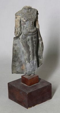
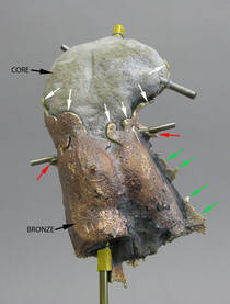
Hollow statues, by definition, were filled with a refractory material, or core, at the time they were . Generally, the core was removed after casting, but in some cases, part (or even all) of the original core may remain, and this material may be analyzed both chemically and petrographically. In the absence of any remnants of original core, there is still potential to find significant clues as to the nature of the core, either by visual examination of the inner surface or by radiography (see I.1§1.1.3, I.1§2.1).
The most common core materials are either clay based (mostly clay, with sand and organic temper; see fig. 10), sand based (mostly sand, bound with clay and possibly organic temper added; see fig. 6), or plaster based (normally gypsum, with or without clay, sand, or organic temper; see fig. 57). Clay-containing bricks may be encountered as well (fig. 8). In clay-based and plaster cores, the clay component may contain raw clay or pre-fired and ground clay (grog), perhaps in the form of ground ceramic or brick. Sand (silica) may be naturally present in clay-based cores, or may be added separately; the extent of silica addition may be so great that refactory material commonly thought of as clay based may be more properly considered sand based.1 For a more detailed discussion of core materials and fabrication, see GI§2.1.1. Casting core studies have often focused on clay-based materials, whereas plaster-based and sand-based cores have been less well studied.2
1.2 Why investigate cores?
The materials used and the method of forming the core are critical parts of the fabrication process, and reflect important technical choices on the part of the . Careful study of residual core can help characterize these materials and methods, which can in turn answer important questions about a bronze sculpture. The analysis of casting cores has been included in the technical study of bronze sculpture for more than thirty years.3 Particularly in the last twenty years, a large number of publications have clearly demonstrated that core analysis is a powerful tool for understanding and characterizing the production methods of bronze sculpture.4
1.2.1 Geographic provenance
Analysis of core material may yield important clues regarding where a bronze sculpture was cast. Specific mineralogical or chemical signatures identified in the raw materials can be compared to databases of geological reference materials, which may establish a likely region of origin. Since foundries have historically tended to choose locally available materials, this may help to geolocate the casting site.
From a methodological point of view, analytical techniques developed for the study of ancient ceramics have been widely applied to core characterization and have influenced the overall approaches taken to the study of cores.5 As a consequence, questions of geographic provenance have been a major focus of many casting core studies (see Case Study 5),6 with chemical compositions used particularly to determine the geographic source of the raw materials. Special attention has focused on the relative amounts of different rare earth elements (REEs),7 though recent studies indicate that existing reference databases should be carefully evaluated before use.8 Shape, texture, and mineralogy of non-clay minerals have also been used for geographic provenancing, albeit less frequently.
1.2.2 Attribution
Core analysis may also shed light on temporal, geographic, or artistic attributions for bronze sculpture. If characteristic practices and raw materials can be identified and associated with particular workshops or artists, it may be possible to support or refute specific attributional hypotheses.
Recently, there has been increased interest in the characterization of workshops and/or related technical practices through characterization of core material (see Case Study 5).9 The characteristics of raw materials (clay, temper, et cetera) may be used to identify workshop practices and recipes employed by specific foundries, in certain periods, to overcome specific technical challenges of bronze casting. Questions may then be raised about the relationships between artists and foundries, and thus, casting core studies can provide another avenue to investigate issues of authenticity.
2 How to investigate cores
The analytical techniques used to study cores fall into two main categories: chemical and petrographic. Chemical analysis generally focuses on the elemental composition of the materials, including their bulk elemental composition and the concentration of certain minor or trace elements that are useful for geographic provenancing. Petrographic analysis focuses on the nature, morphology, and proportion of the different minerals and other additives that are in the core, be they natural or added by the founders (for instance added sand in clay). As much as possible, we recommend combining chemical data and petrographic investigation results to achieve a comprehensive characterization and documentation of the core material.
2.1 Chemical analysis
Chemical analysis follows similar protocols as those described for metals (see II.5), although metal analysis can sometimes be performed in situ. For core analysis it is necessary to obtain samples. Because core material is typically not visible from the exterior, permission for core sampling may be more easily obtained than for metal sampling.
2.1.1 Inductively coupled plasma with atomic (or
optical) emission spectroscopy (ICP-AES or ICP-OES) or
mass spectroscopy (ICP-MS)
Inductively coupled plasma (ICP) refers to a type of high-intensity energy source (torch) that efficiently breaks sample material into individual atoms and then ionizes the atoms. Once atomized and ionized, the total elemental composition can be determined, usually either by atomic emission spectroscopy (ICP-AES) or by mass spectroscopy (ICP-MS).10 AES determines elemental composition by analyzing spectral features of the light produced when the sample material is exposed to the ICP torch. Mass spectrometers are complex instruments and made in several different configurations, but all collect the sample ions and directly measure the mass and abundance of each ion type with a high degree of precision.
ICP analytical techniques are very sensitive to trace quantities of elemental components, often able to detect even a few ppm depending on the detection method. This allows precise quantification of a large number of trace elements, and in particular of rare earth elements,11 which can be extremely useful when addressing provenance questions. Both ICP-AES and ICP-MS involve long, complex, and mandatory chemical preparation of the sample prior to analysis. The analyst must select and follow a dissolution procedure (acid digestion) that is compatible with the sample material and then inject the solution into the plasma torch. MS instruments are generally more sensitive and precise than AES instruments, though this varies somewhat, element by element. MS systems are also significantly more expensive to purchase and difficult to operate.
An alternative to dissolution methods is the use of laser ablation (LA). LA-ICP-MS or LA-ICP-AES uses a focused, pulsed laser to ablate sample material into a plume that is drawn into the ICP instrument directly. Laser spot diameters often fall in the range of 10–300 µm, which can complicate analysis when working with microscopically heterogeneous materials. On the other hand, the precision of the technique has been used to advantage in the analysis of individual mineral grains to trace geographic sources of ceramic material.12 A disadvantage of LA-ICP is that various matrix effects can complicate quantitative analysis.13
The complexity of all ICP variants means that analysis is best performed by private or university laboratories specializing in this technique, and with staff experienced in cultural heritage applications. At the time of publication, costs per sample range from around US$100 to $350 for academic clients.
2.1.2 Neutron activation analysis (NAA)
Neutron activation analysis (NAA) is based on the principle that if a sample material is bombarded with relatively low-energy “thermal” neutrons (usually produced at a nuclear reactor), the atoms in the sample will capture neutrons into their nuclei, forming radioactive isotopes. As these isotopes decay over a period of days or weeks, they emit gamma rays characteristic of each element present in the sample. The gamma rays may be detected and identified, leading to a quantitative estimate of the sample’s total elemental composition. The high penetrating power of neutrons and gamma rays means that NAA provides information from the interior as well as the exterior of the sample material.
Neutron activation is a highly sensitive method for chemical analysis. Data for a wide range of elements, including rare earth elements, can be obtained with sensitivity at the low- and even sub-ppm level. No specific preparation of the sample is needed. However, the restricted availability and cost of this nuclear method makes it more and more difficult to perform. Reliable results require about 50–100 mg of sample material, more than is sometimes possible to obtain from a bronze core. Detection limits for some elements are higher than for ICP-MS, and the analysis requires more time to complete than with other techniques. For these reasons, neutron activation analyses of casting core materials are scarce in the literature. Nonetheless, it remains a very effective tool for geographic provenancing through chemical fingerprinting, and many of the relevant compositional databases have been built using this technique.14
2.1.3 Scanning electron microscopy (SEM)
In scanning electron microscopy (SEM), a sample is placed into a vacuum or near-vacuum and bombarded with a highly focused beam of electrons that is continuously rastered, or scanned, across the surface. Multiple detectors are placed inside the chamber that instantaneously detect different types of emissions from the surface of the sample as the beam is scanning.
For the visualization of core and metal samples, a backscattered electron (BSE) detector is commonly used, which generates an image in which heavy elements (of high atomic number, such as lead) appear bright, while light elements (of low atomic number, such as silicon) appear dark. The precise focus and control of the electron beam allows for the extremely high magnification that SEM is known for.
For elemental analysis, SEM may be coupled with X-ray detectors for energy-dispersive spectroscopy (SEM-EDS or SEM-EDX) or wavelength-dispersive spectroscopy (SEM-WDS or microprobe). These detectors yield X-ray spectra that are useful for elemental analysis.15 Bulk composition can be obtained as well as spot analysis of individual grains or features. Only elemental information is provided, not information about the mineral structure of the components. Both techniques are capable of quantitative elemental analysis, although WDS offers substantially higher precision of measurement than EDS. This puts SEM-EDS at a disadvantage for the purposes of geographic provenancing. The accuracy of both methods may be adversely affected by substantial in the sample.
Today, many major museum and academic laboratories are equipped with SEM-EDS. These instruments may be very valuable for petrographic analysis (see II.7§2.2 below), but for high-precision quantitative elemental analysis, other (unfortunately less accessible) methods such as ICP, NAA, particle-induced X-ray emission spectroscopy (PIXE), or SEM-WDS may be preferred.
2.1.4 X-ray fluorescence analysis (XRF)
X-ray fluorescence analysis (XRF) analysis is a relatively accessible analytical method employed to characterize elemental composition. It may be used to analyze casting core materials16 or bulk metal composition (see II.5§2.1). XRF directs a beam of X-rays at the sample material to stimulate the emission of additional X-rays with specific energies characteristic of the elements present in the sample. As with SEM, the emitted X-rays are detected either with an EDS detector (ED-XRF) or a WDS detector (WD-XRF).
ED-XRF is far more common and is used in all of the portable and handheld XRF instruments that are now widely available in museum and university laboratories. ED-XRF can provide a quick qualitative assessment of the type of core material present. In order to optimize results for light elements such as silicon, aluminum, and calcium, the voltage on the spectrometer should be kept relatively low (around 15 kV), filters should be removed, and helium flush or vacuum should be used if available. Comparison to reference materials (prepared by the research team) may be helpful for general characterization of the core type.
Due to the complex interactions between X-rays and the sample material, quantitative analysis with ED-XRF requires complex calibration procedures and sample preparation that are not well developed for core analysis. In any case, with ED-XRF, sensitivity to REEs will generally not be adequate for geographic provenancing, with the smallest detectable amounts usually in the low hundreds of ppm. Portable ED-XRF instruments now cost between about US$25,000 and $50,000. Commercial laboratories may be able to provide analysis for US$50 or less per sample.
WD-XRF offers significantly higher spectral resolution and sensitivity than ED-XRF, but is less common and much more expensive to own and operate. These instruments reside primarily in commercial and research laboratories focusing on earth sciences. WD-XRF instruments offer the possibility of more rigorous quantitative analysis of core material with sensitivity sufficient to be useful for geographic provenancing, though careful sample preparation is required. The cost per sample is normally less than US$100.
2.1.5 Particle- (or proton-) induced X-ray emission
(PIXE)
PIXE commonly utilizes a highly collimated beam of protons to excite X-ray emission from a sample (other charged particles may be used as well, but protons are by far the most frequently used). It is possible but not necessary for the sample to be placed in a vacuum chamber or flushed with helium to improve detection of light elements. X-ray spectra are collected, usually by an EDS detector, for processing into a quantitative elemental composition. PIXE offers a good balance between analytical performance level and amount of time required for analysis.17 The technique can detect very small quantities of many materials typically present in casting cores, often as little as a few tens of ppm. In the case of point analysis, no specific preparation is required other than placing the sample between two thin polymer films to ease the positioning.
In addition, recent developments in PIXE elemental mapping now permit obtaining a spatial distribution of elements of interest within the sample.18 In this case, the sample should be embedded in resin and polished. Unfortunately, few facilities provide access to ion beam analysis techniques.
2.1.6 X-ray diffraction (XRD)
X-ray diffraction (XRD) takes advantage of the fact that crystalline materials diffract X-rays in predictable and characteristic ways. A collimated beam of X-rays is passed through the sample material, and a detector registers the angles and intensities of the diffracted radiation. Comparing with reference databases allows a determination of the specific crystalline phases present in the sample material.
With respect to core analysis, XRD allows the identification and quantification of important crystalline phases (such as quartz, feldspars, clays, different hydration states of gypsum, et cetera) in clay-, sand-, and plaster-based cores.19 Note that the accuracy of the phase percentages is generally limited to about ±2 wt% for conventional XRD.20 Synchrotron XRD can offer a more precise alternative by using energy ranges allowing for increased accuracy in detection and quantification.
Both conventional and synchrotron XRD can be used to analyze both macro (approx. 100 µm) and micro (less than 50 µm) samples. For macro-scale samples, depending on the geometry of the diffractometer, the sample may be a fine, homogeneous powder mounted on a sample holder or between two Mylar sheets, or a whole fragment of material. In case of micro scale setup, the sample may be placed into a glass capillary or constituted of single crystals or groups of crystallites. In every case, identifying and quantifying mineral phase can help address questions of provenance by comparison with reference material. In case of plaster-based material, the identification of anhydrite (type and proportion) can yield valuable clues about the process of preparing the core.
2.2 Petrographic analysis
Petrographic analysis is ideally done on prepared thin-section samples of intact casting core fragments.21 It is intended to provide information on the different proportions of materials present (clay, plaster, sand, organic temper), the size and shape (morphology) of the particles, the amount and distribution of porosity, and the distribution of different mineral phases). These features are essential for precise characterization of the core.
Some qualitative petrographic studies have been undertaken, but quantitative approaches using point counting or image analysis have provided more significant data and are generally considered indispensable. Quantitative analysis allows efficient identification of clay sources according to the grain size distribution of the natural sand (quartz) components of different clays. Quantitative measures can also help determine whether some portion of the sand temper was intentionally added to clay-rich material because both the size and shape of the quartz particles will likely fall into two distinct categories, as is often the case in ancient ceramics.22
2.2.1 Optical microscopy (OM)
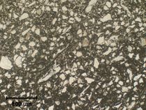
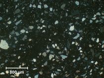
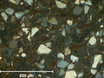
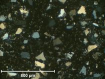
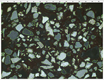
Basic petrographic analysis involves performing optical microscopy (OM) observations on transparent thin sections, usually about 30 µm thick, and preferably at least 1 × 1 cm. Commercial petrographic services will prepare appropriate thin sections from bulk samples for between US $30 and $75. Samples are observed primarily in transmitted light—either plane (linearly) polarized light (fig. 425) or crossed polarized light (figs. 426, 427, 428, see also Case Study 5), with magnifications generally in the 40–400X range, depending on the size of the mineral grains present. Sample material is always placed on a rotating microscope stage so that it can be observed at different angles with respect to the polarization angle of the transmitted light. An experienced petrographer can identify many specific minerals and other inclusions based on observation alone. To identify metallic minerals (which are not transparent), reflected light is also often added.
An optical microscope equipped with polarizing lenses is easy to use, relatively low cost, and available in many laboratories, but the expertise required for competent petrographic examination is substantial, and most researchers of bronze sculpture send their samples to specialists for analysis.
2.2.2 Cathodoluminescence (CL) microscopy
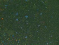
Cathodoluminescence (CL) microscopy is based on the principle that when certain minerals are bombarded by electrons in a vacuum, they emit light of different colors.23 Among these materials, the most commonly encountered in cores are feldspars, which yield a blue luminescence (fig. 429). CL microscopy is often performed in scanning electron microscopes that have been equipped with a CL detector, but optical microscopes can also be adapted with a CL stage.
2.2.3 Scanning electron microscopy (SEM)
SEM can be used for high-magnification observation of petrographic structure. The morphology of organic temper components in loose or crushed sample material may be studied in detail using a secondary electron (SE) detector. This can allow the identification of charcoal, straw, fibers, et cetera. At very high magnification, the morphology of clay grains may be used to identify the types of clay present.
Switching to the BSE detector (see section 2.1.3 above) and working with a polished thin section can yield high-resolution images that show the size and distribution of mineral grains based on their density. These may be used for image analysis (see section 2.2.4 below). Using an EDS detector (see section 2.1.3 above) enables the elemental characterization of mineral grains. Most SEM software is now capable of producing elemental maps that show the distribution of different elements in the sample, as well as phase maps that identify regions with similar elemental compositions.
2.2.4 Image analysis
Quantitative data can be obtained from OM, CL, and SEM images by performing image analysis (fig. 430, see Case Study 5). We recommend taking into consideration a sufficient surface area of the sample (at least a few square millimeters) in order to obtain representative data. At least three different areas of the sample should be analyzed. An alternative is to analyze the entire thin section using a high-resolution scan.24 A dedicated image analysis program may be provided with the microscope, or stand-alone comprehensive image analysis packages can be purchased. Free and open-access software is also available, including ImageJ, which offers good solutions for the automated determination of many petrographic parameters.25
Some important parameters to examine include the relative areas of the different components (including porosity); distribution of the area ratio occupied by each grain with regard to the total area occupied by sandy temper (area-to-area sum);26 the grain size distribution of the different components; and the shape of the grains (including measures of sphericity and angularity).
While much of the image analysis process can now be automated, an experienced petrographer can make crucial judgments about its implementation. For instance, fragments of crushed rock are difficult to delineate, as they are composed of multiple smaller grains, so these should be removed from the automation and processed manually.
3 Sampling and sample preparation
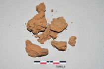
Sampling and sample preparation are of great importance for preserving all the material evidence of the fabrication process. Whereas chemical analysis can be performed on powdery material, it is preferable to work with a coherent block of core, as it enables full characterization of the petrographic structure (fig. 431), chemical heterogeneity, and porosity. Petrographic characterization is ideally performed on a substantial thin section, made from a block of one cubic centimeter or larger if possible. Smaller samples of even a few square millimeters may still provide valuable information. The location of each sample should be clearly documented for future reference.
Cores may be layered structures, so it is important to document the orientation of the sample material as it is removed, and if possible have the thin section prepared as a cross section (with the outer, metal-side surface on one edge and the innermost portion of the sample at the opposite side). In addition, the portion of the core that was in direct contact with the molten metal at the time of casting will have been exposed to much higher temperatures than material slightly removed from the metal surface. This elevated temperature may induce phase transitions in the core material, so it is important to be aware of the orientation of the sample.
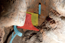
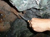
The sampling strategy depends on the degree and locations of access to the core. If there is easy access through an open underside or through casting flaws, for example, burins or chisels may be used to remove core material, which may be hard and compacted (fig. 432). If there is no obvious core material within easy view, exploration with a borescope may reveal pockets of hidden core. If access is difficult, a steel rod may be bent and sharpened into a customized sampling tool specifically shaped to reach the core location. Sampling may be monitored using the borescope, and dislodged blocks may be retrieved with a flexible claw pickup tool. Images collected during the process will ensure that the orientation of the block is documented.
Sometimes only loose, powdery sample material is accessible. For example when a very porous, friable core has partially disintegrated in an archaeological bronze over time, it may be impossible to extract a block sample. Chemical analysis may still proceed with powdery sample material, and so may other types of analysis, such as particle size distribution and shape analysis, provided enough representative sample material is available.
In rare cases, bronzes that allow no access to their cores have been drilled and core extracted through a small hole. The metal drillings have been used for elemental analysis and the core drillings for chemical analysis. Of course, in such cases the knowledge that might be gained must be carefully weighed against the consequent damage,27 and this approach will often be considered inappropriate.
Extracting core material for analysis from a bronze does not affect the long-term stability of the object, as the core only plays a role during the casting process.
4 Risks of misidentification/misinterpretation
Perhaps the greatest risk of misinterpretation lies in the possibility that the material sampled is not original core. In archaeological bronzes there is ample opportunity for soil and sediment to either imitate or contaminate core material. Also, it is not uncommon for cast-in repairs to be made, adding additional core material after the original flawed casting was complete and the original core removed. This may be to repair original casting flaws (executed at the time of the original manufacture) or to repair damages later in the life of the object. While core intended for repairs is, of course, interesting in its own right, it may not be the same as the original core, and should not be confused with it.
If original core is clearly present, care should be taken to extract a sample that appears representative of the bulk of the core. If the core appears to be heterogeneous, then multiple samples may be warranted.
It should also be noted that the filling of hollow bronze sculptures with plaster, long after their original production, is not unheard of. The motives for such treatment are not always apparent, but clearly it happens and may cause much confusion. Common sense is usually the best approach to determine whether the plaster is original or not.28
Casting core studies still suffer from nonstandardized procedures. They are often incomplete and may not be comparable with other aspects of technical examination of bronze statues due to lack of expert data interpretation.
Notes
-
Liu, Siran, Kai Wang, Quanfa Cai, and Jianli Chen. 2013. “Microscopic Study of Chinese Bronze Casting Moulds from the Eastern Zhou Period.” Journal of Archaeological Science 40 (5): 2402–14. https://doi.org/10.1016/j.jas.2012.11.010.. ↩︎
-
Stone, Richard E. 1981. “Antico and the Development of Bronze Casting in Italy at the End of the Quattrocento.” Metropolitan Museum Journal 16:87–116.. To date, unpublished studies of plaster cores include several samples taken from eighteenth-century French bronzes at the C2RMF. Forty-four plaster cores have been examined at the J. Paul Getty Museum, only some of which have been published (Fogelman, Peggy, and Peter Fusco. 2002. Italian and Spanish Sculpture: Catalogue of the J. Paul Getty Museum Collection. Los Angeles: J. Paul Getty Museum. http://www.getty.edu/publications/virtuallibrary/0892366893.html.; Bennett, Shelley M., and Carolyn Sargentson. 2008. French Art of the Eighteenth Century at the Huntington. San Marino, CA: Huntington Library; New Haven, CT: Yale University Press.; Schmidtling II, Ronald C. 2008. “Core Analysis.” In Jane Bassett, The Craftsman Revealed: Adriaen de Vries, Sculptor in Bronze, 35–44. Los Angeles: Getty Conservation Institute. http://www.getty.edu/publications/virtuallibrary/9780892369195.html.; Bassett, Jane, and Francesca G. Bewer. 2014. “The Cut-Back Core Process in Late 17th- and 18th-Century French Bronzes.” In French Bronze Sculpture: Materials and Techniques 16th–18th Century, edited by David Bourgarit, Jane Bassett, Francesca G. Bewer, Geneviève Bresc-Bautier, Philippe Malgouyres, and Guilhem Scherf, 205–14. Paris: Archetype.). core material from four foundries as well as eighteen sand casting cores have been examined at the J. Paul Getty Museum, only two of which have been published (Schmidtling II, Ronald C. 2008. “Core Analysis.” In Jane Bassett, The Craftsman Revealed: Adriaen de Vries, Sculptor in Bronze, 35–44. Los Angeles: Getty Conservation Institute. http://www.getty.edu/publications/virtuallibrary/9780892369195.html.). ↩︎
-
Early published analyses include Stone, Richard E. 1981. “Antico and the Development of Bronze Casting in Italy at the End of the Quattrocento.” Metropolitan Museum Journal 16:87–116.; Milam, Billie, Chandra L. Reedy, and Carol Sussman. 1988. Technical Analysis of Renaissance Bronzes for Provenance Studies: Pilot Project. Malibu, CA: Getty Conservation Institute. http://hdl.handle.net/10020/gci_pubs/tech_analysis_renaissance_bronzes.; Reedy, Chandra L. 1991b. “Petrographic Analysis of Casting Core Materials for Provenancing Studies of Copper Alloy Sculptures.” Archaeomaterials 5:121–63.; and Formigli, Edilberto. 1993. “Antiche terre di fusione.” In Antiche officine del bronzo: materiali, strumenti, tecniche: atti del Seminario di studi ed esperimenti, Murlo, 26–31 luglio 1991, edited by Edilberto Formigli, 69–98. Siena, Italy: Nuova Immagine.. ↩︎
-
Reedy, Chandra L. 1997. Himalayan Bronzes: Technology, Style and Choices. Newark, DE: University of Delaware; London and Cranbury, NJ: Associated University Presses.; Woodward Jr., Hiram W. 1997. The Sacred Sculpture of Thailand: The Alexander B. Griswold Collection. Baltimore: Walters Art Gallery.; Lombardi, Gianni, and Massimo Vidale. 1998. “From the Shell to Its Content: The Casting Cores of the Two Bronze Statues from Riace (Calabria, Italy).” Journal of Archaeological Science 25:1055–66.; Bewer, Francesca G. 2001. “The Sculpture of Adriaen de Vries: A Technical Study.” In Small Bronzes in the Renaissance, edited by Debra Pincus, 158–93. Studies in the History of Art 62. Washington, DC: National Gallery of Art; New Haven, CT: Yale University Press.; Lombardi, Gianni. 2002. “A Petrographic Study of the Casting Core of the Lupa Capitolina Bronze Sculpture (Rome, Italy) and Identification of Its Provenance.” Archaeometry 44 (4): 601–12.; Weisman, Billie Milam, and Chandra L. Reedy. 2002. “Technical Studies on Renaissance Bronzes.” In Materials Issues in Art and Archaeology VI: Symposium Held November 26–30, 2001, Boston, Massachusetts, USA, edited by Pamela B. Vandiver, Martha Goodway, and Jennifer L. Mass, 483–95. Materials Research Society Symposium Proceedings 712. Warrendale, PA: Materials Research Society.; Schmidtling II, Ronald C. 2008. “Core Analysis.” In Jane Bassett, The Craftsman Revealed: Adriaen de Vries, Sculptor in Bronze, 35–44. Los Angeles: Getty Conservation Institute. http://www.getty.edu/publications/virtuallibrary/9780892369195.html.; Lombardi, Gianni. 2009. “The Casting Core Composition and Provenance of the Goljamata Kosmatka (Bulgaria) Bronze Head.” Journal of Archaeological Science 36 (2): 520–27.; Carò, Federico. 2010. “Petrographic and Mineralogical Analysis of the Casting Core of a Chinese Bronze Buddha Maitreya.” Metropolitan Museum Journal 1:155–61.; Mille, Benoît, I. Gajda, F. Demange, C. Pariselle, Y. Coquinot, E. Porto, O. Tavoso, and Antoine Zink. 2010. “Hawtar’athat, Fils de Radaw’il Du Lignage de Shalalum. Une grande statue de bronze du Royaume de Saba’ (Yémen).” Monuments et mémoires de la Fondation Eugène Piot 89: 5–68. https://doi.org/10.3406/piot.2010.1726.; Vincent, B. 2014. “Searching for the Bronze Workshops of Angkorian Cambodia: Petrographic Study Applied to Casting Cores of 11th–12th C. Khmer Bronzes” (unpublished report). Washington, DC: Freer Gallery of Art and Arthur M Sackler Gallery.. ↩︎
-
Waksman, Yona. 2014. “Etudes de Provenance de Céramiques.” In Circulation et provenance es matériaux dans les sociétés anciennes, edited by Philippe Dillmann and Ludovic Bellot-Gurlet, 195–216. Sciences Archéologiques. Paris: Editions des archives contemporaines.; Léon, Yoanna. 2014. “La circulation des savoir-faire techniques des céramiques.” In Circulation et provenance des matériaux dans les sociétés anciennes, edited by Philippe Dillmann and Ludovic Bellot-Gurlet, 225–36. Sciences Archéologiques. Paris: Editions des archives contemporaines.. ↩︎
-
Holmes, Lore L., and Garman Harbotte. 1991. “Provenance Study of Cores from Chinese Bronze Vessels.” Archeomaterials 5 (2): 165–84.; Reedy, Chandra L. 1991b. “Petrographic Analysis of Casting Core Materials for Provenancing Studies of Copper Alloy Sculptures.” Archaeomaterials 5:121–63.; Lombardi, Gianni, and Massimo Vidale. 1998. “From the Shell to Its Content: The Casting Cores of the Two Bronze Statues from Riace (Calabria, Italy).” Journal of Archaeological Science 25:1055–66.; Mugnaini, Sonia, Marco Giamello, Anastasia Pisani, and Salvatore Siano. 2014. “Casting Cores Used to Craft Large Bronze Masterpieces of the Florentine Renaissance and Mannerism.” Journal of Archaeological Science 47 (July): 85–98.. ↩︎
-
For an in-depth review of analytical methods applied to rare earth element analysis see Zawisza, Beata, Katarzyna Pytlakowska, Barbara Feist, Marzena Polowniak, Andrzej Kita, and Rafal Sitko. 2011. “Determination of Rare Earth Elements by Spectroscopic Techniques: A Review.” Journal of Analytical Atomic Spectrometry 26 (12): 2373–90.. ↩︎
-
Hancock, R. G. V., K. Michelaki, W. C. Mahaney, and S. Aufreiter. 2019. “Justification for Reassessing Elemental Analysis Data of Ceramics, Sediments and Lithics Using Rare Earth Element Concentrations and Ratios.” Archaeometry 61 (6): 1430–45.. ↩︎
-
Mugnaini, Sonia, Marco Giamello, Anastasia Pisani, and Salvatore Siano. 2014. “Casting Cores Used to Craft Large Bronze Masterpieces of the Florentine Renaissance and Mannerism.” Journal of Archaeological Science 47 (July): 85–98.; Castelle, Manon, Yvan Coquinot, and David Bourgarit. 2016. “Casting Cores of French Bronze Statues of the 16th and 17th Centuries: Identification of Regional Practices and Artistic Choices.” Microchemical Journal 126:121–31.. However, as early as the 1990s Edilberto Formigli already questioned the fabrication process of the core and its integration within the sculpture’s overall fabrication process (Formigli, Edilberto. 1993. “Antiche terre di fusione.” In Antiche officine del bronzo: materiali, strumenti, tecniche: atti del Seminario di studi ed esperimenti, Murlo, 26–31 luglio 1991, edited by Edilberto Formigli, 69–98. Siena, Italy: Nuova Immagine.). ↩︎
-
Lombardi, Gianni, and Massimo Vidale. 1998. “From the Shell to Its Content: The Casting Cores of the Two Bronze Statues from Riace (Calabria, Italy).” Journal of Archaeological Science 25:1055–66.; Gratuze, B., M. Blet-Lemarquand, and J.-N. Barrandon. 2001. “Mass Spectrometry with Laser Sampling: A New Tool to Characterize Archaeological Materials.” Journal of Radioanalytical and Nuclear Chemistry 247 (3): 645–56.; Goemaere, Éric, Denis Henrotay, Olivier Collette, Mark Golitko, Thomas Delbey, and Thierry Leduc. 2014. “Caractérisation de la céramique médiévale d’Autelbas (Arlon, Belgique) et identification de la source de la matière première.” ArchéoSciences, no. 38 (December): 31–47.; Mugnaini, Sonia, Marco Giamello, Anastasia Pisani, and Salvatore Siano. 2014. “Casting Cores Used to Craft Large Bronze Masterpieces of the Florentine Renaissance and Mannerism.” Journal of Archaeological Science 47 (July): 85–98.. ↩︎
-
Mugnaini, Sonia, Marco Giamello, Anastasia Pisani, and Salvatore Siano. 2014. “Casting Cores Used to Craft Large Bronze Masterpieces of the Florentine Renaissance and Mannerism.” Journal of Archaeological Science 47 (July): 85–98.. ↩︎
-
Gehres, B., and G. Querré. 2018. “New Applications of LA–ICP–MS for Sourcing Archaeological Ceramics: Microanalysis of Inclusions as Fingerprints of Their Origin.” Archaeometry 60 (4): 750–63.. ↩︎
-
Sylvester, Paul. 2008. “Matrix Effects in Laser Ablation-ICP-MS.” In Laser Ablation-ICP-MS in the Earth Sciences: Current Practices and Outstanding Issues, edited by Paul Sylvester, Pap/Cdr edition, 67–78. Quebec: Mineralogical Association of Canada.. ↩︎
-
Holmes, Lore L., and Garman Harbotte. 1991. “Provenance Study of Cores from Chinese Bronze Vessels.” Archeomaterials 5 (2): 165–84.; Reedy, Chandra L. 1997. Himalayan Bronzes: Technology, Style and Choices. Newark, DE: University of Delaware; London and Cranbury, NJ: Associated University Presses.; Lombardi, Gianni, and Massimo Vidale. 1998. “From the Shell to Its Content: The Casting Cores of the Two Bronze Statues from Riace (Calabria, Italy).” Journal of Archaeological Science 25:1055–66.. ↩︎
-
Lombardi, Gianni. 2009. “The Casting Core Composition and Provenance of the Goljamata Kosmatka (Bulgaria) Bronze Head.” Journal of Archaeological Science 36 (2): 520–27.; Mugnaini, Sonia, Marco Giamello, Anastasia Pisani, and Salvatore Siano. 2014. “Casting Cores Used to Craft Large Bronze Masterpieces of the Florentine Renaissance and Mannerism.” Journal of Archaeological Science 47 (July): 85–98.; Vincent, B. 2014. “Searching for the Bronze Workshops of Angkorian Cambodia: Petrographic Study Applied to Casting Cores of 11th–12th C. Khmer Bronzes” (unpublished report). Washington, DC: Freer Gallery of Art and Arthur M Sackler Gallery.. ↩︎
-
Smith, A., H. Botha, F. C. de Beer, and E. Ferg. 2011. “The Examination, Analysis and Conservation of a Bronze Egyptian Horus Statuette.” Nuclear Instruments and Methods in Physics Research Section A: Accelerators, Spectrometers, Detectors and Associated Equipment 651 (1): 221–28.. ↩︎
-
Calligaro, Thomas, Jean-Claude Dran, Evanthia Ioannidou, Brice Moignard, Laurent Pichon, and Joseph Salomon. 2000. “Development of an External Beam Nuclear Microprobe on the AGLAE Facility of the Louvre Museum.” Nuclear Instruments and Methods in Physics Research Section B 161/162/163:328–33.; Dran, Jean-Claude, Thomas Calligaro, and Joseph Salomon. 2000. “Particle-Induced X-Ray Emission.” In Modern Analytical Methods in Art and Archaeology, edited by Enrico Ciliberto and Giuseppe Spoto, 135–66. New York: Wiley-Interscience.; Calligaro, Thomas, Yvan Coquinot, Laurent Pichon, and B. Moignard. 2011. “Advances in Elemental Imaging of Rocks Using the AGLAE External Microbeam.” Nuclear Instruments and Methods in Physics Research Section B-Beam Interactions with Materials and Atoms 269 (20): 2364–72.; Castelle, Manon, Yvan Coquinot, and David Bourgarit. 2016. “Casting Cores of French Bronze Statues of the 16th and 17th Centuries: Identification of Regional Practices and Artistic Choices.” Microchemical Journal 126:121–31.. ↩︎
-
Pichon, L., B. Moignard, Q. Lemasson, C. Pacheco, and P. Walter. 2014. “Development of a Multi-Detector and a Systematic Imaging System on the AGLAE External Beam.” “The 13th International Conference on Particle Induced X-ray Emission (PIXE 2013).” Special issue, Nuclear Instruments and Methods in Physics Research Section B: Beam Interactions with Materials and Atoms 318 (January): 27–31.. ↩︎
-
Stone, Richard E. 1981. “Antico and the Development of Bronze Casting in Italy at the End of the Quattrocento.” Metropolitan Museum Journal 16:87–116.; Holmes, Lore L., and Garman Harbotte. 1991. “Provenance Study of Cores from Chinese Bronze Vessels.” Archeomaterials 5 (2): 165–84.; Lombardi, Gianni, and Massimo Vidale. 1998. “From the Shell to Its Content: The Casting Cores of the Two Bronze Statues from Riace (Calabria, Italy).” Journal of Archaeological Science 25:1055–66.; Schmidtling II, Ronald C. 2008. “Core Analysis.” In Jane Bassett, The Craftsman Revealed: Adriaen de Vries, Sculptor in Bronze, 35–44. Los Angeles: Getty Conservation Institute. http://www.getty.edu/publications/virtuallibrary/9780892369195.html.; Lombardi, Gianni. 2009. “The Casting Core Composition and Provenance of the Goljamata Kosmatka (Bulgaria) Bronze Head.” Journal of Archaeological Science 36 (2): 520–27.; Mugnaini, Sonia, Marco Giamello, Anastasia Pisani, and Salvatore Siano. 2014. “Casting Cores Used to Craft Large Bronze Masterpieces of the Florentine Renaissance and Mannerism.” Journal of Archaeological Science 47 (July): 85–98.. ↩︎
-
Bish, David L., and Jeffrey E. Post, eds. 1989. “Modern Powder Diffraction.” Special issue, Reviews in Mineralogy 20.. ↩︎
-
Lombardi, Gianni, and Massimo Vidale. 1998. “From the Shell to Its Content: The Casting Cores of the Two Bronze Statues from Riace (Calabria, Italy).” Journal of Archaeological Science 25:1055–66.; Newman, Richard. 2006. “Analysis of Core and Investment Samples from Some Aquamanilia.” In Lions, Dragons, and Other Beasts: Aquamanilia of the Middle Ages, Vessels for Church and Table, edited by Peter Barnet and Pete Dandridge, 57–63. New Haven, CT: Yale University Press.; Schmidtling II, Ronald C. 2008. “Core Analysis.” In Jane Bassett, The Craftsman Revealed: Adriaen de Vries, Sculptor in Bronze, 35–44. Los Angeles: Getty Conservation Institute. http://www.getty.edu/publications/virtuallibrary/9780892369195.html.; Lombardi, Gianni. 2009. “The Casting Core Composition and Provenance of the Goljamata Kosmatka (Bulgaria) Bronze Head.” Journal of Archaeological Science 36 (2): 520–27.; Mugnaini, Sonia, Marco Giamello, Anastasia Pisani, and Salvatore Siano. 2014. “Casting Cores Used to Craft Large Bronze Masterpieces of the Florentine Renaissance and Mannerism.” Journal of Archaeological Science 47 (July): 85–98.; Vincent, B. 2014. “Searching for the Bronze Workshops of Angkorian Cambodia: Petrographic Study Applied to Casting Cores of 11th–12th C. Khmer Bronzes” (unpublished report). Washington, DC: Freer Gallery of Art and Arthur M Sackler Gallery.. ↩︎
-
Riederer, Josef. 2004. “Thin Section Microscopy Applied to the Study of Archaeological Ceramics.” Hyperfine Interactions 154:143–58.; Velde, Bruce. 2005. “Use of Image Analysis in Determining Multi-Source Ceramic Materials.” In Pottery Manufacturing Processes: Reconstitution and Interpretation: Acts of the XIVth UISPP Congress, University of Liège, Belgium, 2–8 September 2001, edited by Alexandre Livingstone Smith, Dominique Bosquet, and Rémi Martineau, 95–99. BAR International Series 1349. Oxford: Archaeopress.; Reedy, Chandra L. 2006. “Review of Digital Image Analysis of Petrographic Thin Sections in Conservation Research.” Journal of the American Institute for Conservation 45 (2): 127–46.; Livingood, Patrick C., and Ann S. Cordell. 2009. “Point/Counter Point: The Accuracy and Feasibility of Digital Image Techniques in the Analysis of Ceramic Thin Sections.” Journal of Archaeological Science 36 (3): 867–72.; Dal Sasso, Gregorio, Lara Maritan, Sandro Salvatori, Claudio Mazzoli, and Gilberto Artioli. 2014. “Discriminating Pottery Production by Image Analysis: A Case Study of Mesolithic and Neolithic Pottery from Al Khiday (Khartoum, Sudan).” Journal of Archaeological Science 46 (June): 125–43.. ↩︎
-
Chapoulie, Rémy, Béatrice Robert, and Sandrine Casenave. 2016. “The Cathodoluminescence Phenomenon Used for the Study of Ancient Ceramics and Stones.” Cities of Memory: International Journal on Culture and Heritage at Risk 1 (1): 53–72.. ↩︎
-
Reedy, Chandra L., Jenifer Anderson, Terry J. Reedy, and Yimeng Liu. 2014. “Image Analysis in Quantitative Particle Studies of Archaeological Ceramic Thin Sections.” Advances in Archaeological Practice 2 (4): 252–68.. ↩︎
-
Reedy, Chandra L. 2006. “Review of Digital Image Analysis of Petrographic Thin Sections in Conservation Research.” Journal of the American Institute for Conservation 45 (2): 127–46.. ↩︎
-
Velde, Bruce. 2005. “Use of Image Analysis in Determining Multi-Source Ceramic Materials.” In Pottery Manufacturing Processes: Reconstitution and Interpretation: Acts of the XIVth UISPP Congress, University of Liège, Belgium, 2–8 September 2001, edited by Alexandre Livingstone Smith, Dominique Bosquet, and Rémi Martineau, 95–99. BAR International Series 1349. Oxford: Archaeopress.. ↩︎
-
Milam, Billie, Chandra L. Reedy, and Carol Sussman. 1988. Technical Analysis of Renaissance Bronzes for Provenance Studies: Pilot Project. Malibu, CA: Getty Conservation Institute. http://hdl.handle.net/10020/gci_pubs/tech_analysis_renaissance_bronzes.; Weisman, Billie Milam, and Chandra L. Reedy. 2002. “Technical Studies on Renaissance Bronzes.” In Materials Issues in Art and Archaeology VI: Symposium Held November 26–30, 2001, Boston, Massachusetts, USA, edited by Pamela B. Vandiver, Martha Goodway, and Jennifer L. Mass, 483–95. Materials Research Society Symposium Proceedings 712. Warrendale, PA: Materials Research Society.; Reedy, Chandra L. 2006. “Review of Digital Image Analysis of Petrographic Thin Sections in Conservation Research.” Journal of the American Institute for Conservation 45 (2): 127–46.; Reedy, Chandra L., and Pieter Meyers. 2007. “New Methods for Analyzing Thin Sections of Casting Core Materials: A Case Study with Southeast Asian Bronzes.” In Scientific Research on the Sculptural Arts of Asia: Proceedings of the Third Forbes Symposium at the Freer Gallery of Art, edited by Janet G. Douglas, Paul Jett, and John Winter, 103–14. London: Archetype.. ↩︎
-
Neptune (1583) by Barthélemy Prieur (French, 1536–1611) in the Musée de Melun, France (inv. 802, Millet 17), was filled with plaster once removed from its fountain (Seelig-Teuwen, Regina, David Bourgarit, and Francesca G. Bewer. 2014. “Barthélemy Prieur Fondeur, son atelier, ses méthodes de travail.” In French Bronze Sculpture: Materials and Techniques 16th - 18th Century, edited by David Bourgarit, Jane Bassett, Francesca G. Bewer, Geneviève Bresc-Bautier, Phillippe Malgouyres, and Guilhem Scherf, 18–38. London: Archetype.). In Donatello’s (Italian, ca. 1386–1466) Spiritelli in the Musée Jacquemart-André, Paris (inv. MJAP-s1773), nineteenth-century newspaper fragments were found in the plaster core, attesting that the plaster was not original but resulting from a later (Castelle, Manon, Marc Bormand, Yannick Vandenberghe, and David Bourgarit. 2019. “Two of a Kind: Shining New Light on Bronze Spiritelli Attributed to Donatello.” Studies in Conservation 65 (4): 200–211.). ↩︎