Additional Contributors: Jane Bassett, Arlen Heginbotham, Alexis Komenda, Anne Maigret, Ruven Pillay, Yosi Pozeilov
This chapter aims to provide specialists and nonspecialists with insight into a range of photography and 3D modeling techniques for the study of sculpture. Nonspecialists—those who find themselves in situations where professional photographers cannot be present—will find general guidance regarding production of high-quality images and what results can be expected. Advantages and drawbacks of available methods are discussed, including relative capability, costs, and time considerations when possible and relevant. For a synthesis, see table 13 Open viewer.
This chapter provides, by necessity, only abbreviated and general guidelines. For detailed guidance on best practice for photographic documentation of cultural heritage, including image and metadata management, see Warda, Jeffrey, ed. 2011. The AIC Guide to Digital Photography and Conservation Documentation. 2nd ed. Washington, DC: American Institute for Conservation of Historic and Artistic Works., Pozeilov, Yosi A. 2015. Digital Imaging and Documentation for Art Conservation. 4th ed. Edited by Laura Cherry. Digital Photography for Art Conservation. Self-published, Lulu.com., and other references cited below.
1 Visible-light photography: most common techniques
1.1 Lighting a bronze sculpture
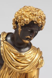
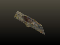
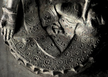
Many bronze sculptures have reflective or glossy surfaces (fig. 293), and particularly in these cases, a uniform and diffuse light will help minimize specular reflection. Diffuse lighting means that light is directed at the subject from large-area sources, usually accomplished by use of reflection umbrellas or diffuser screens in front of the light sources or by using flat-panel LED arrays. Normal flash photography, which involves a single small light source near the camera, is usually undesirable. Normally, at least two light sources are recommended. For documentary photography, shadows should not be too “hard” and there should be more even light than for catalogue or commercial images, where dramatic lighting is often preferred.1
Use lights that are designed for photography and have a high color rendering index (CRI) and always include a standard color and scale chart. For documentary photography, use only one type of illumination source at a time; if light sources of different color temperatures are used (such as window light in combination with photo lamps), the color rendition may be significantly different on different parts of the subject. In most circumstances, it is recommended to use a tripod with the camera set on “aperture priority.” Selection of a high f-stop will significantly improve sharpness and depth of field, but will require longer exposures, which in turn necessitates a tripod. In situations where a high f-stop is not adequate to capture the required depth of field, a series of images may be captured, with the focal point moving stepwise from the nearest to farthest point on the sculpture. So-called focus stacking software has become increasingly available in common image processing programs, allowing the image set to be blended into a single image where all areas appear in focus. A tripod is essential for focus stacking, and all individual images should be archived along with the stacked image.
Particularly with dark sculpture, the full dynamic range (the difference in luminance between the darkest and lightest areas) may not be captured in one exposure. In such cases multiple, “bracketed” images of different exposures should be captured. High dynamic range (HDR) algorithms can composite different exposures into a single image, but always retain the original files.
1.2 Raking-light photography
For the documentation of fine surface topography such as tool marks and evidence of wear, raking-light photography may be useful. Here, instead of diffuse lighting, a single, strongly directional light source is positioned at a very low angle to the surface, typically around 10 degrees. This arrangement causes minor irregularities in surface topography to cast shadows, making them easier to perceive. Altering the direction of the light source can reveal different features on any given surface. Raking light can be useful for standard photography, macro photography, and even photomicrography (see II.2§2.1 below).
1.3 Macro photography
In many circumstances, close-up or macro photography will be necessary to resolve details of technical interest. Specialized macro lenses, which are designed to minimize distortion, are preferred for this purpose. The closer-up the image is, the smaller the depth of field will be, and thus the greater importance of using a high f-stop. In situations where it is desirable to hold the camera by hand, dedicated macro flash units, such as lens-mounted ring flash units, can provide sufficient diffuse or directional light for high speed and high f-stop macro imaging.
1.4 Image quality and resolution
The quality of images produced will depend on both the resolution of the camera’s sensor (fig. 369) and the quality of the lenses used. For this reason, a professional-quality digital camera is recommended. As of the year 2023, a typical mirrorless camera may be equipped with a 8200 × 5500 pixel (45 megapixel) sensor, which will capture a one-meter-high statue with a vertical resolution of 82 pixels/cm, corresponding to a pixel size of 0.12 mm on the statue. Professional medium-format cameras with 100+ megapixel sensors can be of great benefit, but are cost prohibitive for many labs.
1.5 Time required to properly photograph a bronze
The time required to photograph a bronze sculpture in a professional studio can vary dramatically depending on the nature of the object and the number of details required. The simple documentation of six low-gloss statuettes via four or five general views of each might take two and a half days: half a day for the setup, one day for the photographs, and one day for post-processing. On the other hand, the full documentation of a single large, dark, and glossy bronze with numerous details may take more than six days: half a day for the setup, two days for capturing images (which will require several lenses), and four days for post-processing.2
2 Visible-light photography: less common techniques
2.1 Photomicrography
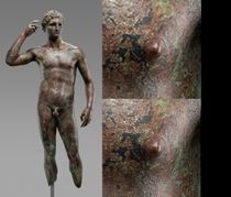
The imaging of surface features substantially smaller than one millimeter (polishing marks, details of tool marks, small inclusions, porosities, excrescences) may require higher magnification than macro photography, namely photomicrography. Traditional stereomicroscopes installed on articulated arms are very useful for this purpose, and these can be fitted with cameras for image capture.
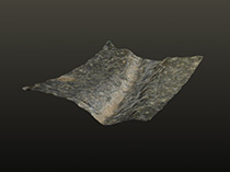
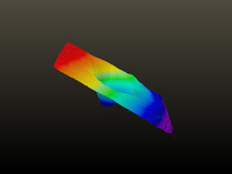
High-quality digital microscopes (without eyepieces) offer additional capabilities and are increasingly widely used for technical examination of bronze sculpture. Most importantly, these microscopes usually incorporate automated focus stacking procedures that overcome the shallow depth of field inherent to photomicrography. Because the focus adjustments are precisely controlled by stepper motors, the microscope’s software can reconstruct a scaled 3D of the field of view. Such topographic reconstructions can be useful for documenting, characterizing, and comparing fine features such as tool marks (figs. 273, 285, 286, see Case Study 4). With this type of 3D model, the sources of errors for precise calculations are numerous and measurements should therefore be considered with caution. Digital microscopes often have polarizers incorporated that can greatly reduce specular reflection, and some can be adapted for use with ultraviolet illumination.
If extremely fine, high-precision topographic measurements are required, a method called microtopography may be useful. Various techniques are available, with resolutions as small as 0.1 µm. This technique is expensive, slow, and can generally be used only on samples smaller than 20 cm across.3
There are also a wide range of low-cost handheld digital microscopes available with a variety of magnification ranges that can be tethered directly to a laptop computer. Some models are equipped with ultraviolet and/or polarized light sources. While the image quality is generally not as good as more expensive laboratory- and research-grade equipment, they may be adequate for routine examination.
2.2 Endoscopy
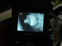
Endoscopes (alternately called borescopes) are common tools for those studying the techniques of bronze sculpture. They are rigid or flexible tubes with a lens at one end and some optical or digital means to transmit an image through the lens to an eyepiece, camera, or digital sensor. In most cases, a light source is incorporated into the endoscope’s tip. They can be used to examine the interior of a hollow sculpture by inserting the boroscope into the base (or any aperture) (figs. 38, 370). Image quality tends to be low, and images generated by a boroscope tend to be difficult to interpret because there is no external frame of reference, so careful attention to image annotation and captioning is critical. Recording video with audio narrative can also be an effective method of documentation.
The cost of endoscopes can vary tremendously. Models produced for industry or the medical field that have articulated heads (whose direction can be controlled from the exterior) and software that enables making measurements may be priced in the tens of thousands of US dollars, while simple USB models are available for less than one hundred US dollars.
2.3 Reflectance transformation imaging
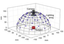
Reflectance transformation imaging (RTI) can be of great utility in documenting and visualizing fine surface topography as well as color and gloss. In this method, both the object and the camera are fixed; only the point light source (typically a flash unit) moves. A large number of photographs are taken of the object (usually between twenty-four and one hundred) as the light source is systematically moved around the object (fig. 371). A reflective sphere is placed into the frame of each image and the position of the specular reflection of the light source on the surface of the sphere is used to calculate the direction of the light source. The software for compositing and viewing RTI images is open-source and free, making the technique readily accessible (fig. 241). Several mathematical image enhancement features are available that allow interactive relighting and enhancement of color and surface shape attributes.4
3 Imaging with nonvisible light
3.1 Ultraviolet fluorescence photography
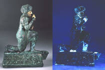
Organic materials such as varnish, pigments, glue, resins, and a variety of restoration materials may fluoresce visible light when exposed to (invisible) ultraviolet light (UV). Photography of UV-induced visible fluorescence (often misleadingly called UV photography for short) may thus prove useful to visualize such materials on the surface of a sculpture (fig. 191). The method may be used in conjunction with macro photography and photomicrography.
Several types of UV light sources are available, including LEDs, fluorescent tubes, and arc lamps. Generally, lamps emitting at 365 nm with minimal visible light emission are preferred. Standard digital cameras can be used for UV fluorescence photography, but require special filters in front of the lens to block UV and infrared (IR) transmission to the camera’s sensor. For details see Pozeilov, Yosi A. 2015. Digital Imaging and Documentation for Art Conservation. 4th ed. Edited by Laura Cherry. Digital Photography for Art Conservation. Self-published, Lulu.com..
Risks of misidentification/misinterpretation
Although some materials emit important and specific color under UV, a variety of heterogeneous materials may emit similar fluorescence. Moreover, fluorescence is also affected by long-term light exposure and thermal aging. As a consequence, fluorescence does not generally allow for precise material characterization. Once fluorescent materials are localized by UV examination and photography, complementary analysis may be required to identify the materials present (see II.5).
Additionally, organic may be present even if little or no fluorescence is detected. Over time, copper ions may migrate into organic coatings, and this can cause dramatic quenching of fluorescence. In addition, some organic materials possibly present on bronze sculpture, including many synthetic resins, do not fluoresce.
3.2 Thermography and infrared (IR) photography
Repair , , and other discontinuous areas of a bronze are often thermally isolated from their surrounding regions. When the surface is heated, for example by the sun or a tungsten lamp, the heat will be kept longer there, dissipating more slowly than on the surrounding large surfaces. IR thermography (IRT) typically uses a camera sensitive to long-wave IR light (about 9000–14,000 nm) to image heat transfer and buildup on an object’s surface. The thermograms thus obtained have been used on bronze sculpture to investigate and map , mechanical and , as well as inlays.5 Thermography in the mid-IR (about 3000–5000 nm) has also been applied to cultural heritage.6 Imaging in the near-IR (about 700–10.1 µm) or short-wave IR (1100–2500 nm), commonly used on many other types of artwork, is generally of limited utility in the examination of bronze sculpture.
4 Color measurement
Color measurement has been used on occasion for research related to bronze sculpture, either to evaluate conservation treatment methods,7 to characterize the color of different alloys,8 or to characterize the color of .9 Most of these applications have been made in the context of experimental simulation (see II.9§1.2), where sample size, uniformity, and geometry can be controlled.10
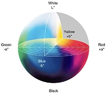
A colorimeter or spectrophotometer may be used to measure color. These instruments illuminate a sample area under controlled conditions and measure the light reflected by the object. Colorimeters use filters to measure the amount of red, green, and blue light reflected from the sample, while spectrophotometers generate a detailed spectrum of the reflected light. In both cases, the resulting color measurements are normally made using the CIELAB color space, defined by three variables, L*, a*, and b*, as defined by the Commission Internationale de l’Éclairage (CIE) (fig. 372).11 Color measurement instruments commonly used for museum objects are usually handheld and cost anywhere from several hundred to several thousand US dollars.
Color measurement of bronze patinas can also be accomplished using visual matching to standard color swatches such as Munsell soil color charts as described in Devogelaere, Jonathan. 2017. “The Colour Palette of Antique Bronzes: An Experimental Archaeology Project.” Experimental Archaeology 2017 (2). https://exarc.net/ark:/88735/10289..12
Risks of misidentification/misinterpretation
The science behind color generation, color perception, and color measurement is complex. This is particularly true in the context of bronze sculpture because the color of bare metal is generated through a completely different physical mechanism than the color of patinas and oxidation layers.13 Bronze sculpture is thus much impacted by goniochromism—that is, the change of color with the angle of the observer.14 Where patinas (either organic, inorganic, or mixed) are not entirely opaque, meaningful and reproducible color measurement by any method may be difficult to achieve. Recently, new methods have been proposed that characterize surface appearance (particularly of patinas and gilding layers) using more complex procedures that take into account color, gloss, and translucency at several viewing angles.15
Color measurement is theoretically possible with digital photography following a color calibration protocol. However, in practice, color measurement of bronze sculpture with photography is extremely challenging. In addition to the difficulties mentioned above, accurate color measurement requires that the light falling on the measured area be the same color and intensity as the light falling on the calibration reference card and have the same directionality. This poses a practical problem with three-dimensional sculpture, since any change in the angle of the surface, as well as local inconsistencies in intensity or color temperature of the lighting, can affect the measured color.
5 Photogrammetry and 3D scanning
Photogrammetry and 3D scanning can produce three-dimensional renderings of objects useful for documentation and dissemination of information on the appearance of a sculpture. There are currently three main techniques available: photogrammetry, structured light scanning, and laser scanning. These can be combined in various ways to produce mixed models. All of these technologies are developing rapidly, and costs for both hardware and software are falling. In addition to recording appearance, these methods can also be useful for physical measurement; this latter aspect of their use is addressed in II.4§2.2.2 and II.4§2.3.4. All of these methods require specialized knowledge and training, though photogrammetry is probably the easiest to learn and has the additional advantage of not requiring specialized hardware.
5.1 Photogrammetry
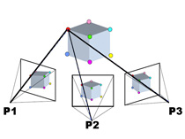
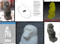
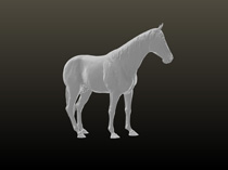
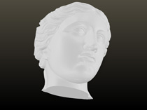
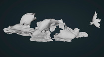
In photogrammetry, many photographs are taken of the subject from different angles and a 3D model is derived from a computational analysis of the individual images using dedicated software (fig. 373). Most photogrammetric work done in cultural heritage has been carried out with professional-grade digital cameras, though it is also possible to build very high resolution models using medium-format cameras that record images of 100 megapixels and higher. Applications for smartphones are also now also available at lower resolution.
Depending on the quality and resolution of the source images, photogrammetry can provide a quite high-fidelity model of a bronze sculpture with good color accuracy, particularly if the photography is done in a studio with controlled lighting. These images can be useful for both public and scholarly audiences, providing the opportunity for high-quality web-based interactive viewing. Photogrammetry can also be carried out using UV and IR photography.16 Photogrammetry is being used more and more for bronze sculpture, notably to design models (fig. 374) and mounts (fig. 375), or to test reassembly of fragmentary statues. Large-scale bronzes can be captured using scanners mounted to drones.17

5.2 Structured light scanners
Structured light scanners shine a regular pattern of light (usually a series of parallel lines) onto an object and then record an image of the pattern with a camera that is offset from the projection source. Software then analyzes the apparent distortion of the light patterns by the three-dimensional surface and generates a computer model of the form. Most structured light scanners also record standard photographic images of the subject using onboard flash lighting, which alternates at high speed with structured light pulses.
The lenses and sensors used in structured light scanners are generally of lower quality than a professional digital camera, and lighting and color management are often a challenge.18 As a result, structured light scans often yield a final model that is less realistic and detailed in appearance than a photogrammetric model (figs. 376, 377), though the dimensional accuracy may be as good or superior. It may thus be used for precise measurements (fig. 378, see II.4§2.2.2).
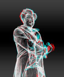
5.3 Laser scanners
Most laser scanners used in cultural heritage employ a rapidly rotating mirror to deflect a visible or invisible laser beam in a pattern that sweeps across the subject. The scanner integrates a range finder that accurately measures the distance to each of the millions of points that the beam traces, building up a high-resolution 3D model of the form. Often multiple scans from different vantage points are combined to produce the final model (fig. 379). Most laser scanners integrate cameras to provide “texture,” or photographic detail, that can be overlaid onto the 3D model (figs. 380, 381). However, as with structured light scanning, the relatively low quality of the cameras used can result in models that are less realistic and detailed in appearance than a good photogrammetric scan.
Notes
-
Yosi A. Pozeilov recommends that the main light point toward the object at an angle of 50–60 degrees. The second lamp should be at 35–45 degrees. For this and other pointers, see Pozeilov, Yosi A. 2015. Digital Imaging and Documentation for Art Conservation. 4th ed. Edited by Laura Cherry. Digital Photography for Art Conservation. Self-published, Lulu.com., 88. ↩︎
-
These data were kindly provided by two photographers at the C2RMF, Anne Maigret and Alexis Komenda. ↩︎
-
Mélard, Nicolas, Clotilde Boust, Gabrielle Cogné, and Anne Maigret. 2016. “Comparison of Imaging Techniques Used in the Microanalysis of Paleolithic Mobiliary Art.” Journal of Archaeological Science: Reports 10 (December): 903–9.; Page, M. J., C. Boust, N. Mélard, D. Robcis, and G. Obein. 2016. “3D Surface Acquisition: Comparison of Two Microtopographic Equipments When Measuring Materials of Cultural Heritage.” In Proceedings of the 4th CIE Expert Symposium on Colour and Visual Appearance: 6–7 September 2016, Prague, Czech Republic, edited by Commission internationale de l’éclairage, 111–20. Vienna: CIE.. ↩︎
-
Dellepiane, Matteo, Massimiliano Corsini, Marco Callieri, and Roberto Scopigno. 2006. “High Quality PTM Acquisition: Reflection Transformation Imaging for Large Objects.” In CIPA / VAST / EG / EuroMed 2006: 37th CIPA International Workshop dedicated on e-Documentation and Standardisation in Cultural Heritage: incorporating: the 7th International Symposium on Virtual Reality, Archaeology and Intelligent Cultural Heritage, the 4th EUROGRAPHICS Workshop on Graphics and Cultural Heritage, 1st Euro-Med Conference on IT in Cultural Heritage, Nicosia, Cyprus, October 30-November 4, 2006, edited by Marinos Ioannides, David B. Arnold, Franco Niccolucci, and Katerina Mania, 179–86. Aire-la-Ville, Switzerland: Eurographics Association. http://dx.doi.org/10.2312/VAST/VAST06/179-186. Free software and viewers can be downloaded from the Cultural Heritage Imaging website: http://culturalheritageimaging.org/Technologies/RTI/. ↩︎
-
Orazi, Noemi, Fulvio Mercuri, Ugo Zammit, Stefano Paoloni, Massimo Marinelli, Augusto Giuffredi, and Carlo Stefano Salerno. 2016. “Thermographic Analysis of Bronze Sculptures.” Studies in Conservation 61 (4): 236–44.; Mercuri, Fulvio, Noemi Orazi, Ugo Zammit, Augusto Giuffredi, Carlo Stefano Salerno, Cristina Cicero, and Stefano Paoloni. 2017. “The Manufacturing Process of the Capitoline She Wolf: A Thermographic Method for the Investigation of Repairs and Casting Faults.” Journal of Archaeological Science: Reports 14:199–207.; Mercuri, F., G. Caruso, N. Orazi, U. Zammit, C. Cicero, O. Colacicchi Alessandri, M. Ferretti, and S. Paoloni. 2018. “Interface Thermal Conductance Characterization by Infrared Thermography: A Tool for the Study of Insertions in Bronze Ancient Statuary.” Infrared Physics and Technology 90 (May): 31–39.. Different profiles, and consequently techniques of mechanical repairs, are detectable on the Capitoline Wolf. ↩︎
-
See Gavrilov, D., R. Gr. Maev, and D. P. Almond. 2014. “A Review of Imaging Methods in Analysis of Works of Art: Thermographic Imaging Method in Art Analysis.” Canadian Journal of Physics 92 (4): 341–64.. ↩︎
-
Heginbotham, Arlen, Vincent Beltran, Briana Feston, Ruven Pillay, and Christina Simms. 2014. “An Evaluation of Protective Coatings for Brass in Indoor Environments, with an Emphasis on Boulle Marquetry.” In Furniture Finishes: Past, Present and Future of Transparent Wood Coatings, edited by Miko Vasques Dias, 84–103. Amsterdam: Stichting Ebenist.; Letardi, Paola, Barbara Salvadori, Monica Galeotti, Andrea Cagnini, Simone Porcinai, Alessandra Santagostino Barbone, and Antonio Sansonetti. 2016. “An In Situ Multi-Analytical Approach in the Restoration of Bronze Artefacts.” Microchemical Journal 125:151–58.. ↩︎
-
Devogelaere, Jonathan. 2017. “The Colour Palette of Antique Bronzes: An Experimental Archaeology Project.” Experimental Archaeology 2017 (2). https://exarc.net/ark:/88735/10289.; Mödlinger, Marinne, Maikel H. G. Kuijpers, Dennis Braekmans, and Daniel Berger. 2017. “Quantitative Comparisons of the Color of CuAs, CuSn, CuNi, and CuSb Alloys.” Journal of Archaeological Science 88:14–23.; Radivojević, M., J. Pendić, A. Srejić, M. Korać, C. Davey, A. Benzonelli, M. Martinón-Torres, N. Jovanović, and Z. Kamberović. 2018. “Experimental Design of the Cu-As-Sn Ternary Colour Diagram.” Journal of Archaeological Science 90:106–19.. ↩︎
-
Benzonelli, A., I. C. Freestone, and M. Martinón-Torres. 2017. “A Better Shade of Black: Effects of Manufacturing Parameters on the Development of Ancient Black Bronzes.” Archaeometry 59 (6): 1034–49.; Formigli, Edilberto. 2013a. Colore e luce nella statuaria antica in bronzo. Rome: L’Erma di Bretschneider.. ↩︎
-
Letardi, Paola, Barbara Salvadori, Monica Galeotti, Andrea Cagnini, Simone Porcinai, Alessandra Santagostino Barbone, and Antonio Sansonetti. 2016. “An In Situ Multi-Analytical Approach in the Restoration of Bronze Artefacts.” Microchemical Journal 125:151–58. carried out direct measurements on statues. Devogelaere, Jonathan. 2017. “The Colour Palette of Antique Bronzes: An Experimental Archaeology Project.” Experimental Archaeology 2017 (2). https://exarc.net/ark:/88735/10289. focused on ancient bronzes but working on experimental coupons. See also Formigli, Edilberto. 2013a. Colore e luce nella statuaria antica in bronzo. Rome: L’Erma di Bretschneider.. ↩︎
-
For an accessible synthesis of color science in the context of cultural heritage see Berns, Roy S. 2016. Color Science and the Visual Arts: A Guide for Conservators, Curators, and the Curious. Los Angeles: Getty Trust Publications.. For more in-depth scientific insights see Hunt, R. W. G, and Michael R. Pointer. 2011. Measuring Colour. 4th ed. Hoboken, NJ: Wiley. and Johnston-Feller, Ruth. 2001. Color Science in the Examination of Museum Objects: Nondestructive Procedures. Tools for Conservation. Los Angeles: Getty Conservation Institute. http://www.getty.edu/conservation/publications_resources/pdf_publications/pdf/color_science.pdf.. And for a deep dive into color models and their application see Fairchild, Mark D. 2013. Color Appearance Models. Chichester, UK: Wiley.. ↩︎
-
Devogelaere, Jonathan. 2017. “The Colour Palette of Antique Bronzes: An Experimental Archaeology Project.” Experimental Archaeology 2017 (2). https://exarc.net/ark:/88735/10289.; Munsell Color. 2000. Munsell Soil Color Charts. New York: Munsell Color.. ↩︎
-
Nassau, Kurt. 1987. “The Fifteen Causes of Color: The Physics and Chemistry of Color.” Color Research & Application 12 (1): 4–26.. ↩︎
-
The term was first proposed in McCamy, C. S. 1996. “Observation and Measurement of the Appearance of Metallic Materials. Part I. Macro Appearance.” Colour Research and Application 21 (4): 292–304.. ↩︎
-
Arteaga, Yoko, Clotilde Boust, Angèle Dequier, and Jon Yngve Hardeberg. 2021. “Image-Based Goniometric Appearance Characterisation of Bronze Patinas.” Color and Imaging Conference 29 (1): 294–99. https://doi.org/10.2352/issn.2169-2629.2021.29.294.; Arteaga, Yoko, Diane Marchioni, Stephanie Courtier, Clotilde Boust, and Jon Y. Hardeberg. 2022. “Appearance-Based Evaluation of Varnish Removal Methods in Gilded Surfaces.” Preprint. In Review. https://doi.org/10.21203/rs.3.rs-1933665/v1.. ↩︎
-
Lanteri, Luca, Giorgia Agresti, and Claudia Pelosi. 2019. “A New Practical Approach for 3D Documentation in Ultraviolet Fluorescence and Infrared Reflectography of Polychromatic Sculptures as Fundamental Step in Restoration.” Heritage 2 (1): 207–15.. ↩︎
-
For more, see http://culturalheritageimaging.org/Technologies/Photogrammetry/. ↩︎
-
Felicísimo, Ángel M., and María-Eugenia Polo. 2022. “Measurement and Control of Colour Fidelity in Scanned 3D Models for Heritage Conservation.” Journal of Cultural Heritage 56 (July): 159–66. https://doi.org/10.1016/j.culher.2022.06.010.. ↩︎