In 2014, when the Harvard Art Museums (HAM) agreed to participate in the APPEAR project, analytical findings about the Romano-Egyptian funerary portraits in the care of the museums and new questions emanating from the research became a rich resource in teaching undergraduate and graduate courses in art history, chemistry, ancient studies, and beyond. Students of all disciplines are fascinated by these alluring fragmentary depictions and by the collaborative research of the curatorial, conservation, and scientific departments at the museums. What we know about these portraits and how we come to know it can be shared not just with students or specialist groups but with any audience we welcome through our doors; with this in mind we launched our exhibition, Funerary Portraits from Roman Egypt: Facing Forward (August 27–December 30, 2022). In the following, we give a brief overview of the exhibition and its goals, and then focus on a particular topic that was explored in the display: What can the scientific study of materials and techniques contribute to the identification of individual painters or workshops?
Exhibition
The exhibition centered around the profound connection the ancient portraits evoke in modern viewers, the scientific investigations undertaken to better understand the painting techniques, and our responsibility, in stewarding these fragments, to honor the memory of the long-dead people depicted in the portraits by acknowledging the desecration of their burials. The show was team-curated by Susanne Ebbinghaus, curator of ancient art; Georgina Rayner, conservation scientist; Kate Smith, paintings conservator; and Jen Thum, Egyptologist and museum educator.1 In a new model of co-curation for the Harvard Art Museums, we each brought a particular lens to the work, enriching the truly collaborative presentation.
In a welcome departure from our usual practices, we invited students and faculty, along with Harvard community members of Egyptian heritage and identity, to join us for Community Conversations designed to shape the exhibition early in the process. As a result, we changed our minds about some things—for example, we had already intuited that using the word “mummy” to refer to a person was a form of objectification (many other museums are moving away from this term now, too) and thought we might say “mummified remains,” but Harvard community members of Egyptian heritage asked us to avoid the word “remains” as well. We also heard helpful suggestions about how to frame the exhibition for people not very familiar with ancient Egyptian culture, such as the inclusion of a label describing the funerary rituals surrounding the portraits’ use. These conversations helped us think more clearly about how to discuss provenance, identity, and the troubling legacy of the modern “mummy” paint in the gallery space, both in label texts and in gallery talks and virtual events.
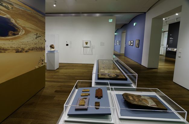
The exhibition’s introductory room (fig. 17.1) was dedicated to contextualizing the portraits by addressing what they are, their cultural functions, and their paths into the museum. Our curators’ statement at the start of the show reminded viewers to keep the sensitive and problematic nature of these collections in mind. In the absence of actual mummified bodies, we borrowed the burial shroud of Tasheret-Horudja from the nearby Museum of Fine Arts, Boston (MFA 54.993), to contextualize the portraits and suggest human scale. We also borrowed inscribed wooden “mummy labels” from the MFA and the Brooklyn Museum (BKM) to present visitors with the identities and personhood of real ancient people—not those depicted in the portraits on view, but others (such as Plenis and Hor, who are only known today through their labels)—to show how those buried in this manner were meant to be remembered, as people, with bodies, likenesses, and names.2
The second, central space (fig. 17.2) focused on eight funerary portraits (five from the HAM collection joined by three loans), our analytical investigation, and what new understanding was gleaned from the materials we identified. An electromagnetic spectrum displayed with a corresponding set of technical images derived from infrared through radiographic bandwidths illustrated the various imaging techniques and what they can tell us. Brief video loops near the portraits presented technical imaging and pigment analysis results, inviting the viewer to join us as we investigated each painting.
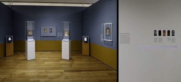
We showed two of the eight panel portraits in the round so that both sides of the portraits could tell their stories of ancient construction and modern dismantling. The back of an encaustic portrait of a woman (HAM 1923.60) retains linen wrappings and resins from the funerary equipment, a stark and intimate reminder of the panel’s original placement above a deceased woman’s face. The back of a tempera portrait of another woman (HAM 1939.111) reveals a modern restoration of the perimeter, as well as stamps and labels that permit at least a partial reconstruction of its provenance.
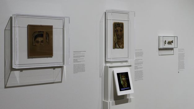
We took a new approach to what was conveyed by the labels accompanying the objects in the exhibition, providing more specific information than usual. The media line was expanded from the basic description into a rich cataloguing of identified materials, from the wood species of the panel to individual pigments used in the paint mixtures. We also included what is known of the portraits’ provenance, their modern history after they were taken from the burial context. Most museum visitors are unfamiliar with the concept of provenance. The information offered in HAM’s online database is difficult to parse, comprising short phrases separated by semicolons, with terms that may themselves be unfamiliar (what, for example, is a bequest?). By stringing the information available for each object into a cohesive and easy-to-follow narrative, we gave visitors the opportunity to better understand the trajectory that each object took before entering the museum where it now resides. Anecdotal evidence suggests that these efforts were well received.
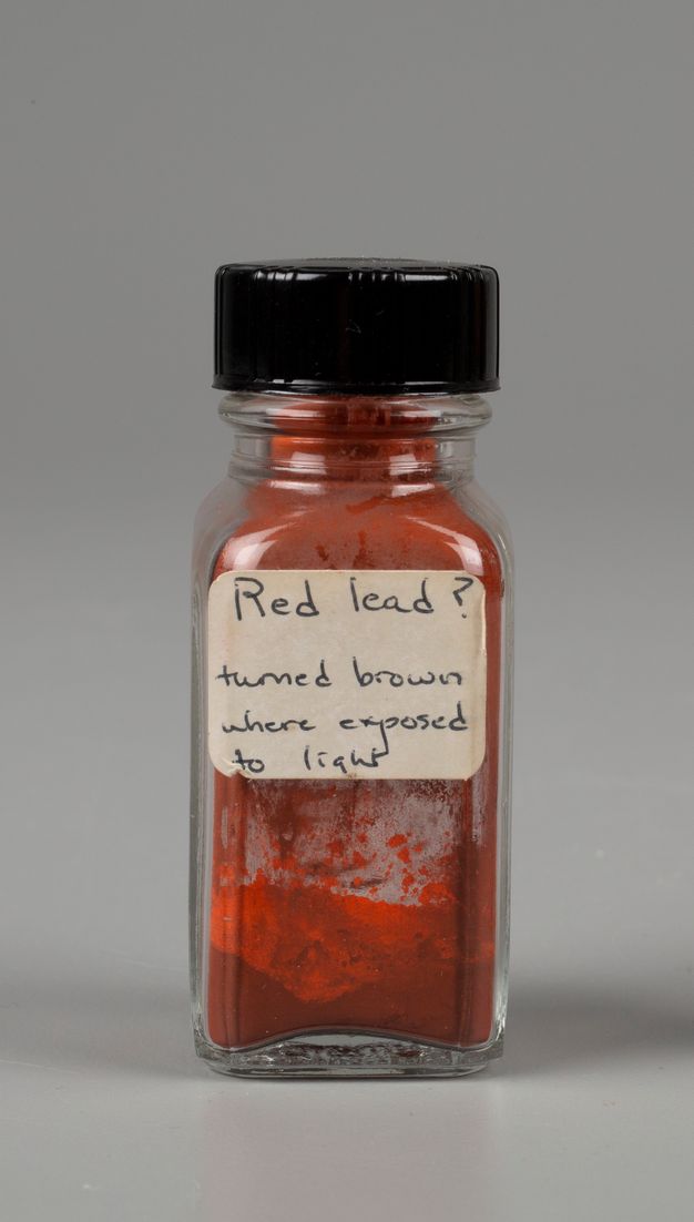
We confronted the damage and desecration inherent in collecting these portraits by showing fragmentary portraits along with early twentieth-century tubes of the brown paint known as “mummy” (fig. 17.3). Valued by artists for centuries for its handling properties and transparent brown tone, the powdered pigment for this oil paint was made from mummified ancient Egyptian individuals.3 Edward Forbes, second director of the Harvard Art Museums, collected two tubes from the London artist supplier Charles Robeson in 1915 for his renowned pigment collection.
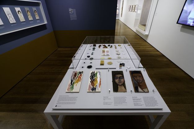
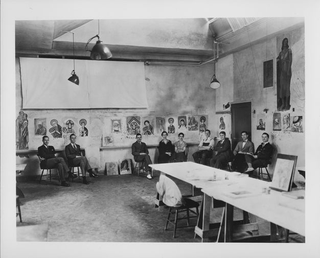
The portrait subjects were not the only ancient people we engaged with while studying the paintings. In identifying the pigments, binders, and wood panels the portrait painters chose, we felt both the presence and absence of the ancient artists. The last area of the exhibition (fig. 17.4) evoked an artists’ studio. A horizontal, table-like case showed pigments in their plant and mineral sources and as prepared colored powders along with beeswax and animal-skin glue binding media, all of which were used in antiquity. Visitors could touch panels painted with these materials to feel the difference between encaustic and tempera paint. The Harvard Art Museums have always been a place for the study of artworks from a material perspective, where students are taught historical techniques through making copies (fig. 17.5). In 1931, a student named Muriel Mussels chose the fragmentary portrait of a man (1924.80) to copy in an encaustic technique for a research project. Her copy (1938.GNRA.1) has lived in our collection ever since. In preparation for and coinciding with the run of our exhibition, we invited Francisco Benitez, a contemporary encaustic painter who focuses on replicating ancient techniques,4 to offer workshops for students and visitors to try their hand at encaustic painting. A current Harvard student, Namirah Quadir, lent her copy to the exhibition. It was displayed next to the 1931 student copy alongside the artist materials, a testament to a century of learning through making at our teaching museum.
A Workshop Active in Ancient Philadelphia (Fayum)
Three exhibition portrait loans gave us an opportunity to compare a group of panel portraits that had been attributed through visual analysis to the hand of a single artist, or possibly a workshop or “school,” which painted funerary portraits exclusively in a tempera technique (fig. 17.6). The attribution to this artist of the only tempera portrait in HAM’s collection, representing a woman with graying hair in a white tunic (fig. 17.6B), goes back almost a century to the study of German scholar Heinrich Drerup. In 1933 he named the painter after a portrait of a bearded man in the collection of Würzburg University, Germany, which he admired, although he thought that the portrait of a woman now at Harvard was painted in a “rough and careless” manner. Among the characteristics of his “Würzburg Painter,” Drerup noted the “whiplash determination and elegance” of the lines as well as the “flourishing polychromy basking in unbroken colors.”5 Almost fifty years later, David Thompson baptized the same artist or “extensive school” as the “Saint Louis Painter” after a portrait of a woman now residing at the Saint Louis Art Museum (SLAM 128:1951; fig. 17.6A), who looks like an older sister of the woman depicted in the portrait at HAM.6 Most recently, Branko van Oppen de Ruiter assembled a group of more than twenty portraits that he associated with a single workshop based on shared features such as the elongated nose, lip shape, and earrings, as well as the hatching technique used to suggest volume and shading in the flesh tones (fig. 17.7).7 Many portraits in this group are remarkable for showing wrinkles and strands of gray or whitish hair. Such signs of advanced age are comparatively rare among Egyptian funerary portraits overall.8
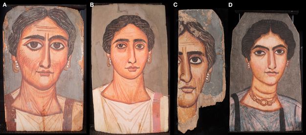
We borrowed the eponymous portrait from SLAM to join the HAM example, along with another, fragmentary painting attributed to the Saint Louis Painter that is now in the care of the Yale University Art Gallery (YUAG 2011.102.1; fig. 17.6C) and a portrait not previously connected with this painter, which now resides at the Smith College Museum of Art (SCMA 1932.9.1; fig. 17.6D). An investigation into the portraits’ provenance, based on museum and other archival records as well as the labels and stamps preserved on the reverse of some of the panels, suggests that all four were part of a cache of over three hundred portraits amassed in the late 1880s by Theodor Graf (1840–1903), an Austrian businessman active in Cairo who also dealt in rugs, papyri, and ancient textiles. Graf stated that the portraits came from er-Rubayat in the northeastern Fayum, and “er-Rubayat” is the location with which the portraits attributed to the Saint Louis Painter have tended to be associated in art historical scholarship. However, a brief report by the Austrian surveyor P. Stadler, who sold portraits to Graf, indicates that while er-Rubayat was the nearest modern settlement, the panels were in fact found closer to the ruins of Philadelphia, some six miles to the east.9 Current excavations in the cemetery area outside this ancient city have brought to light complete as well as fragmentary panel portraits associated with burials in tombs of various architectural forms, including masonry-built catacombs.10
The ongoing (and future) excavations of the cemeteries of ancient Philadelphia hold the promise of providing context for the dissociated and dispersed fragments that were part of Graf’s stock as well as information about the date of the portraits attributed to the Saint Louis Painter. Originally assigned to the fourth century CE based on stylistic traits considered to be late antique, they are now more often thought to reflect Roman hairstyles current in the late second and the beginning of the third centuries CE.11 However, many of the hairstyles in the Egyptian portraits are quite simple and therefore difficult to place. Perhaps they are shaped more by the painting style than by the fashion of the time. Proponents of a later date thought that the portraits were indicative of a switch in the Fayum from the encaustic to the tempera technique, following the assumption that tempera was quicker and easier to use. Even well over a century after the discovery of these portraits, there still is much to discover about them, including a better understanding of which stylistic and technical features are owed to the chosen binding medium, which ones might be a sign of the times (or region, for that matter), and which might be characteristic of a painter or workshop.
Hallmarks of the Workshop
Having several examples of portraits attributed to the Saint Louis Painter in physical proximity at HAM allowed us to closely observe and analytically compare various features. Details such as the earrings and manner of building volume in the faces reveal what appears to be a range of approaches to a common visual goal, implying a workshop environment rather than an individual artist.
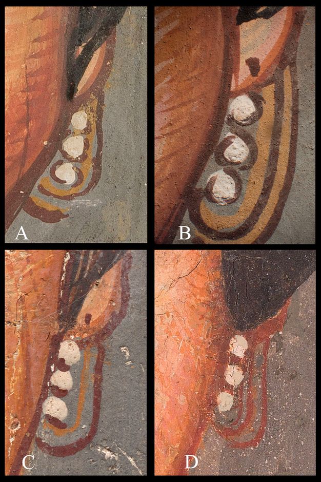
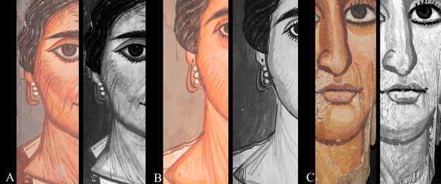
The earrings in each of these four portraits are remarkably alike at first glance (fig. 17.8): gold loops expressed with ochre pigments and decorated with three white beads each. Nine of the eleven female portraits currently proposed as products of this ancient Philadelphia “school” wear these exact earrings. Close inspection of the jewelry reveals a range of finish and detailing from portrait to portrait. The SLAM portrait’s double loops and bead outlines (fig. 17.8A) are described with interrupted lines, and there is no piercing mark on the earlobes. The portrait at HAM (fig. 17.8B) shows piercing marks, and the double loops and bead outlines are painted in continuous lines. In the example at YUAG (fig. 17.8C), there is a single loop each of yellow and red, and piercing, while the beads are simply underlined. And in the SCMA portrait (fig. 17.8D) there are double loops of red ochre only, no piercings are indicated, and the three beads are separated by two red ochre dots between them, rather than surrounded by ochre.
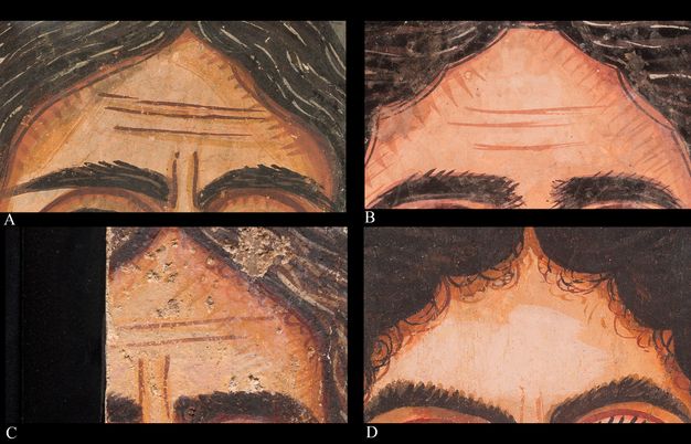
Volume and shadow in the flesh tones are suggested by shaping the face, neck, ears, and nose with bands of paint that narrow in width and darken in color toward the outer contours. Hatch marks often perpendicularly cross these contouring bands either everywhere in the flesh or in particular places like the cheeks, further animating these surfaces. A subtle technique in all but the SCMA example is that the lightest flesh tones were applied earliest, and the contouring details applied last. The SCMA portrait, by contrast, has the highlight tone applied in a final swirl of paint visible at the center of the forehead, with the earliest color field visible in the mid-tone (see fig. 17.7D).
We pursued technical imaging and materials analysis to deepen the investigation of these formal relationships, revealing many shared material features and significant differences.12 We also interrogated panel wood species, ground layers, preparatory sketches, and pigments to establish patterns of material choices and application techniques.13
Wood Species
Three of the four portraits were sampled for wood identification, and a variety of wood types were found: native sycomore fig (Ficus sycomorus) for HAM, willow wood (Salix sp.) for SLAM, and tamarisk (Tamarix aphylla) for SCMA, all native to Egypt. This diversity could suggest that in a wood-scarce region, Egyptian artists and workshops used what materials were at hand or available at a given time.
Priming Layer
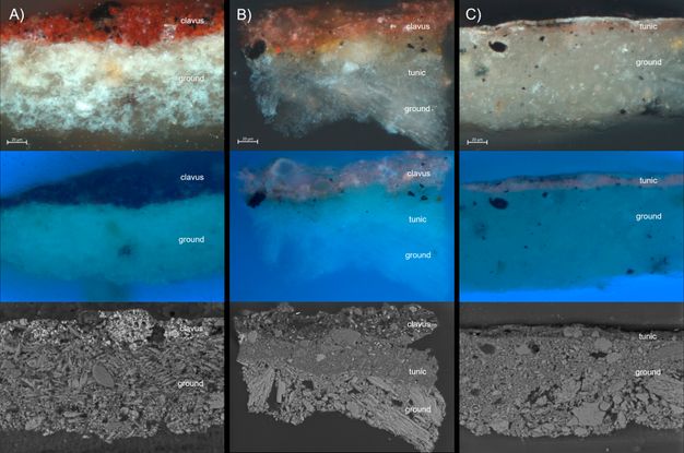
For the tempera painting technique, a priming layer appears to be a typical14 preparation for the wood surface to accept the thin, matte paint produced with animal glue. SLAM and HAM have white calcium sulfate priming, with some ochre inclusions observed in cross sections of minute samples (fig. 17.9). YUAG’s portrait was not sampled, but close visual examination through a microscope paired with elemental information from scanning X-ray fluorescence spectroscopy (XRF) implies a similar method, though with much coarser texture, likely due to a larger pigment particle size than in the priming layers of the other portraits. The SCMA priming layer is gray to the naked eye where visible; in cross section the priming layer is also calcium sulfate but toned with carbon-containing particles. The choice of priming color may have impacted the painting process, lending tone to the final paint layers and affecting the degree of contrast as the design was developed. The color and elemental makeup of the priming may well be a marker of individual painters or methods within a workshop.
Preparatory Sketch
Once the ground layer was in place, a loose preparatory sketch was made in liquid black paint on the SLAM, HAM, and YUAG portraits, with broad strokes visible in infrared reflectography (IR) and in some cases detectable with the naked eye through the thin surface paint (fig. 17.10). No infrared or visible evidence of sketching was detected in the SCMA example. While there may in fact be no preparatory sketch, it is also possible that the carbon content in the ground obscures a carbon-based sketch. It might also be that the artist sketched an initial design with another material not detected with IR.
Paint Stratigraphy
The HAM paint layers are markedly thicker than in the other two sampled portraits (see fig. 17.9) at SLAM and SCMA (fig. 17.11). There is a white layer for the tunic and a pink layer for the clavus (shoulder stripe) over the calcium sulfate ground, and both paint layers are substantial. In the SLAM portrait the white ground was left exposed to stand as the white of the tunic without additional paint, and the clavus was laid directly onto that surface in a single thin layer of red ochre. The SCMA sample has two paint layers over a gray ground: the tunic color followed by the dark stripes describing folds. The two layers together are still thinner than even one of the layers from the portrait at HAM. While the thickness of paint applied can certainly vary among a single artist’s body of work, it seems unlikely that an artist would make economic use of the priming in one portrait but then paint a white tunic over a white ground in another; these differences imply the presence of more than one artist, each accomplishing a similar visual goal with different approaches.
| Layer | Saint Louis Art Museum | Harvard Art Museums | Smith College Museum of Art |
|---|---|---|---|
| Clavus |
About 30 μm thick Red: ochre (iron rich plus clay), trace gypsum, unidentified black (likely carbon) |
About 25 μm thick. Madder, ochre (iron oxide), gypsum, alunite, unidentified black (likely carbon) | |
| Tunic | About 35 μm thick. Alunite* (a mixture of fine grained with some coarse particles), small amounts of gypsum and ochre (iron oxide), unidentified black (likely carbon) |
About 12 μm thick. Madder, possible Al-based mordant, gypsum, possible clay (Al, Si) |
|
| Ground |
At least 95 μm thick. Gypsum (much finer particle size than HAM and SCMA) with possible clay, one particle of alunite much finer ground in comparison. |
At least 60 μm thick. Gypsum (very coarse and variable) |
At least 10.5 μm thick. Gypsum (very coarse but with a higher proportion of fine material compared to HAM), trace ochre (iron oxide and clay), unidentified black (likely carbon) |
Note: Layer thickness varies across the individual layers in the sample. Measurements given in the table above are an average. Cross sections did not capture the wooden panel, so the true thickness of the lowest layer (ground) is unknown.
*Some of the alunite contained jarosite.
Use of Red Lead
In all but the SCMA example, the lips were painted with a lead-based pigment. Lead’s high density makes it visible as a light area in X-radiographs (fig. 17.12), whereas most of the other pigments used in the tempera medium are less dense and therefore penetrated by X-rays, which means that they are less visible in the X-radiograph. The YUAG portrait was not X-radiographed, but its XRF elemental distribution map for lead shows that the lips of the portrait were painted with a lead-containing pigment (fig. 17.12C). In the HAM and SLAM examples (fig. 17.12B and A), red lead is suggested by XRF detection of elemental lead in the context of the presenting red color, although it is not the brilliant color typical of red lead, but instead closely resembles the iron ochre reds used elsewhere in the facial features. In situ Raman spectroscopy of the lips of the HAM portrait, in the X-ray opaque area, conclusively identified a mixture of red lead and hematite (red ochre). However, red lead can discolor to a duller brown color when suspended in a porous, aqueous medium such as animal glue because oxygen can interact with the pigment (fig. 17.13).15 Did the choice of a more expensive, synthetic,16 brilliant red color for the lips have a symbolic significance in its ancient context, or was it simply an aesthetic choice that has altered over time?
Common Features and Outliers
The portraits now residing at SLAM, HAM, and YUAG align in many material ways: all have a similar panel shape, white priming layer, underdrawings, and red lead in the lips. But the SCMA portrait is different in these particulars as well as in formal analysis of its content: the woman portrayed in this painting wears a necklace painted with an arsenic-containing pigment, possibly orpiment or realgar; her neck is described with horizontal instead of diagonal brush strokes; and her tunic is painted with a mixture of indigo and red lake. Are these differences an indication that the SCMA portrait does not belong to this group, despite its visual connections to the other paintings? Or is it part of a subset that could reveal itself through examination of the larger group?
Use of Madder Lake
Ultraviolet-induced visible fluorescence (UVF) images (fig. 17.14) reveal the orange-pink fluorescence characteristic of organic red madder lake in the HAM, YUAG, and SCMA examples. In these portraits, the artist used madder lake in finishing strokes on the surface in a markedly similar way, hatching along the bridge of the nose, eyes, lips, chin, earlobes, and cheeks, as well as in the extant clothing where it is used as the color of the clavus in the HAM example and mixed with indigo for the tunic in the SCMA example. In the SLAM portrait, the result is unclear; red lake may be present in the lower layers of the flesh tones, as a slight fluorescence is faintly visible between surface strokes, but even if the pigment was used on this portrait, it is not applied in the manner so strikingly similar in the other three examples.
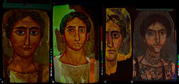
Alunite
Because the grounds of all four portraits were pigmented with calcium sulfate, we anticipated that the white pigment elsewhere in the panels would be the same. However, the HAM portrait is unique within this group; the UVF image suggests a distinction between the ground layer and the white tunic color (see fig. 17.14). The tunic’s white paint has a dull brownish color in UV, while the calcium sulfate ground fluoresces with a different, faintly blue-white color. This separate layer for the tunic can be seen clearly in cross section (see fig. 17.9B) and primarily contains an alunite mineral with similar proportions of sodium and potassium, referred to here as alunite. Alunite was confirmed analytically in samples taken from other areas of the portrait in both the gray background and the pink clavus. While a trace of alunite was identified in the white layer of the SLAM cross section, it does not appear to have been used in any substantial way for that portrait.
Alunite has not been suggested as a typical pigment in the Egyptian palette,17 although recent studies have identified it in a selection of tempera portraits.18 Of particular interest is its identification in the flesh tones, gray background, and white tunic of the portrait of a bearded man19 in the collection of the Martin von Wagner Museum at Würzburg University, mentioned above as a portrait by the artist now commonly referred to as the Saint Louis Painter.
Conclusion
In some respects, the portrait of a woman in our care at HAM is materially similar to the one at YUAG, in others to that at SLAM, and often to neither. The SCMA example, not previously associated with the Saint Louis Painter, bears remarkable visual and material similarities to the other three; for this reason, it should perhaps also be included in this artistic grouping. The formal similarities combined with the range of materials and techniques across these paintings suggest multiple hands working toward a common template or visual type in a workshop setting. The possibility of multiple artists working simultaneously or even sequentially over several generations could also explain the absence or presence of features such as the hatched use of madder lake, the use of alunite as a white pigment, or the application of a gray priming color. Whatever the case, if these paintings were made in a larger workshop environment, we are left with the question of how the faces were rendered so similarly. Was there a template or model of some kind? If so, how was it communicated, and over what period? Our present study sets the groundwork for future technical examinations of panels attributed to the Saint Louis Painter from collections around the world. This will eventually give us a clearer picture of the work of the artists who provided ancient Philadelphians with the portraits required to care for and memorialize the dead.
To date, the group of paintings discussed above has been named after the modern, Western collections where a particular example is held: Würzburg, Germany, or Saint Louis, Missouri. These names have also implied a single painter, while our study suggests the existence of a group of artists working toward a similar visual goal. We propose a new name for this group that emphasizes the original site of creation, rather than the location of the modern collections that benefit from the desecration and fragmentation of the burials that once contained the portraits. The term “Ancient Philadelphia Workshop” lacks sufficient specificity, as another group of paintings attributed to “the Brooklyn Painter” were also made in ancient Philadelphia, but in a markedly different style.20 We therefore propose the name “Ancient Philadelphia Workshop of the Hatch Mark Style,” taking our cue from the long-observed application technique employed across the group. It is our hope that this renaming will be the first of many changes in museums’ approaches to funerary portraits as we seek to consider these remarkable paintings in their original cultural context and aim to pay respect to both those portrayed and those who portrayed them.
Notes
-
The exhibition was accompanied by a digital research tool with the same title; see Ebbinghaus, Susanne, Georgina Rayner, Kate Smith, and Jen Thum. 2022. “Funerary Portraits from Roman Egypt: Facing Forward.” Harvard Art Museums. Accessed September 2, 2022. https://harvardartmuseums.org/tour/770. (https://harvardartmuseums.org/tour/770). ↩︎
-
Thum, Jen. 2022. “The People behind the Portraits.” Harvard Art Museums. https://harvardartmuseums.org/tour/770/stop/2623.. ↩︎
-
Woodcock, Sally. 1996. “Body Colour: The Misuse of Mummy.” The Conservator 20 (1): 87–94.. ↩︎
-
Benitez, Francisco. 2018. “Francisco Benitez.” http://www.franciscobenitez.com/.. ↩︎
-
Drerup, Heinrich. 1933. Die Datierung der Mumienporträts. Ferdinand Schönigh. (with antisemitic stereotypes that were unfortunately rather common at the time). ↩︎
-
Thompson, David L. 1976. The Artists of the Mummy Portraits. J. Paul Getty Museum.; Thompson, David L. 1982. Mummy Portraits in the J. Paul Getty Museum. J. Paul Getty Museum.. ↩︎
-
See van Oppen de Ruiter, this volume. ↩︎
-
Matheson, Susan. 2022. “Individuality and Old Age in the Painted Funerary Portraits of Roman Egypt.” Harvard Art Museums. Accessed December 29, 2024. https://harvardartmuseums.org/tour/770/slide/12398.. ↩︎
-
Ebbinghaus, Susanne, Georgina Rayner, Kate Smith, and Jen Thum. 2022. “Tracing the Path to the Museum.” Harvard Art Museums. Accessed September 2, 2022. https://harvardartmuseums.org/tour/funerary-portraits-from-roman-egypt-facing-forward-2/slide/12413.. ↩︎
-
Gehad, Basem, Lorelai H. Corcoran, Mahmoud Ibrahim, Ahmed Hammad, Mohamed Samah, Abd Allah Abdo, and Omar Fekry. 2022. “Newly Discovered Mummy Portraits from the Necropolis of Ancient Philadelphia–Fayum.” BIFAO 122, 245–64. https://journals.openedition.org/bifao/11727.. ↩︎
-
Borg, Barbara. 1996. Mumienporträts: Chronologie und kulturelle Kontext. Philipp von Zabern.; Borg, Barbara. 1998. “Der zierlichste Anblick der Welt…” In Ägyptische Porträtmumien. Phillip von Zabern.. ↩︎
-
Our colleagues at Yale University and the Saint Louis Art Museum performed extensive analysis on their own portraits in support of this study and they, along with staff at the Smith College Museum of Art, allowed us to do additional imaging prior to the exhibition in order to obtain consistent conditions where possible. ↩︎
-
SLAM infrared reflectography carried out with an Opus Osiris camera InGaAS-array detector, 900–1700 nm operating wavelength.
Technical imaging at HAM was carried out with a Canon Mark III 5D DSLR camera and Zeiss 50mm Makro-Planar ZE lens. The internal camera filtration was removed to allow for full bandwidth response at the detector. External filters and light sources per technique: PECA 918 and Wratten 2E filters with Elinchrom Style RX 1200 strobes for visible and with UV Systems Triple Bright 3 for ultraviolet-induced visible fluorescence photography, Tiffen 87A filter with Lowell Pro tungsten lights for infrared reflected photography, and with Sylvania LED 13 PAR 30LN bulbs for visible-induced infrared luminescence photography. Infrared reflectography for HAM and SCMA was carried out with Xenics Tigris imager (InSb detector) filtered to the 1.5–1.8 micron range. Computed X-radiography was carried out with a Comet MXR-320/26 tube and Carestream Industrex Flex HR detector plate.
A Bruker Artax XRF spectrometer with a Silicon Drift Detector (SDD) and a rhodium anode X-ray tube was used for point analysis. The primary X-ray beam is collimated to give a spot size of 0.65mm. Using the Bruker Artax (version 7.6) software, spectra were acquired for 100 seconds live time at 50kV and 600 μA. A helium flux was used to increase the detection efficiency for light elements (atomic number of potassium and lower).
Elemental distribution maps were generated using a Bruker M6 Jetstream scanning XRF spectrometer equipped with a Rh target and operated at 50 kV and 600 μA. The Yale portrait was imaged using a 200 μm nominal spot size, 150 μm pixel spacing, a dwell time of 50 ms per pixel, and He purge of 0.6 L/min. The data were processed in PyMca and DataMuncher.
Samples for cross-section analysis were embedded in Bio-Plastic liquid polyester casting resin (Ward’s Natural Science). Mounted samples were ground to exposure using a Buehler Handimet 2 roll grinder with Carbimet abrasive paper rolls ranging in grit from 240 to 600. Samples were then polished using a Buehler Metaserv 2000 polisher with 6µm and 1µm Buehler MetaDi Monocrystalline Diamond Suspension.
Cross sections were observed using a Zeiss Axio Imager.M2m upright microscope equipped with four objectives (5x, 10x, 20x, and 50x) and a Zeiss Axiocam 512 Color digital camera. Images were captured using the Zeiss Zen 2.6 (blue edition) software. Visible light and bright field conditions utilized a halogen lamp and either an EPI-polarization filter cube or an EPI-Bright Field cube respectively. Ultraviolet conditions utilized a mercury vapor lamp and either a DAPI filter cube (excitation BP 450–490, beam splitter FT 510 and emission BP 515-565) or a FITC filter cube (excitation BP 450–490, beam splitter FT 510 and emission LP515).
The cross sections were analyzed using a JEOL JSM-IT500LV SEM (tungsten filament) with an Oxford Instruments X-MaxN SDD, 80 mm2 detector (resolution Mn Kα, 126 eV) running the Oxford Instruments AZtec software (version 4.2 SP1). The SEM was operated in low vacuum mode at a chamber pressure of 70 Pa, with an operating voltage of 20 kV, beam current optimized for dead time of analysis and working distance of 10 mm. The cross sections were not coated prior to analysis.
FTIR in transmission mode was performed using a Bruker Vertex 70 infrared bench spectrometer coupled to a Bruker Hyperion 3000 infrared microscope. Samples were compressed onto a diamond cell with a stainless-steel roller prior to analysis. Using the Bruker OPUS (version 6.0) software, spectra were recorded between 4000 and 600 cm-1 at 4cm-1 spectral resolution and 32 scans per spectrum. The collected spectra were compared to in-house and IRUG databases.
Raman analysis was conducted using a Bruker Optics Senterra dispersive Raman microscope with an Olympus BX51M microscope equipped with 20x and 50x long working distance objectives and using the Bruker OPUS (version 7.5) software. The Raman spectrometer has three laser sources, 532 nm, 633 nm, and 785 nm. The optimum laser source depends on the pigment analyzed, but in general, blue and green pigments were predominantly analyzed with the 532 nm laser at 2 mw or 5 mW power and other colors with the 785 nm laser at 10 mW power. Spectra were compared with reference libraries, particularly the RRUFFTM database, using the Opus software.
The following procedures were carried out by Caroline Cartwright in the laboratory of the Department of Scientific Research at the British Museum. Because of the three-dimensional nature of wood anatomy, each wood sample, irrespective of its size, was fractured manually to show transverse, radial longitudinal, and tangential longitudinal sections (TS, RLS, and TLS). Each TS, RLS, and TLS wood section was then mounted onto aluminum stubs. Examination of the wood samples and comparative reference specimens (prepared and mounted using the same method) was undertaken in a variable-pressure scanning electron microscope (VP SEM), Hitachi S-3700N, using the backscattered electron (BSE) detector at 15 kV, with the SEM chamber partially evacuated (40Pa). Magnifications ranged from 35x to 1000x. The preferred working distance was ca. 14 mm but was raised or lowered from 10.6 mm to 16.5 mm (as required). With the BSE detector, 3D mode (rather than Compositional) was preferentially selected for maximum topographical information and to maximize the potential for revealing diagnostic features for identification. Further details on wood identification methods and techniques can be found in Cartwright, Caroline R. 2015. “The Principles, Procedures and Pitfalls in Identifying Archaeological and Historical Wood Samples.” Annals of Botany 116 (1): 1–13. https://doi.org/10.1093/aob/mcv056., Cartwright, Caroline R. 2020. “Understanding Wood Choices for Ancient Panel Painting and Mummy Portraits in the APPEAR Project through Scanning Electron Microscopy.” In Svoboda and Cartwright 2020. https://www.getty.edu/publications/mummyportraits/part-one/2., and Cartwright in this volume. ↩︎
-
“APPEAR Ancient Panel Painting Examination, Analysis, and Research Database,” J. Paul Getty Trust, 2022: https://www.appeardatabase.org/login. Sorting the database entries by binding medium and reviewing all tempera portraits, we find most employ a priming between the wood substrate and the paint layer. ↩︎
-
FitzHugh, Elisabeth West. 1986. “Red Lead and Minium.” In vol. 1 of Artists’ Pigments: A Handbook of Their History and Characteristics, edited by Robert Feller. National Gallery of Art.. ↩︎
-
Walton, Marc S., and Karen Trentelman. 2009. “Romano-Egyptian Red Lead Pigment: A Subsidiary Commodity of Spanish Silver Mining and Refinement.” Archaeometry 51 (5): 845–60.. ↩︎
-
Lee, Lorna, and Stephen Quirke. 2000. “Painting Materials.” In Nicholson and Shaw 2000.; Scott, David A. 2016. “A Review of Ancient Egyptian Pigments and Cosmetics.” Studies in Conservation 61 (4): 185–202. https://doi.org/10.1179/2047058414Y.0000000162.. ↩︎
-
Dietemann, Patrick, Heike Stege, Ursula Baumer, Andrea Obermeier, Christoph Steuer, Luise Sand, Catharine Blӓndsdorf, Elisabeth Fugmann, and Kristian Kaiser. 2017. “Pigmente und Bindemittel antiker Mumienporträts.” In Inkarnat und Signifikanz: Das menschliche Abbild in der Tafelmalerei von 200 bis 1250 im Mittelmeerraum, edited by Yvonne Schmuhl and Esther Pia Wipfler. Zentralinstitut für Kunstgeschichte.; Freccero, Agneta. 2000. Fayum Portraits: Documentation and Scientific Analysis of Mummy Portraits Belonging to Nationalmuseum in Stockholm. Acta Universitatis Gothoborgensis.; Brøns et al., this volume; and Verri et al., this volume. ↩︎
-
Dietemann, Patrick, Heike Stege, Ursula Baumer, Andrea Obermeier, Christoph Steuer, Luise Sand, Catharine Blӓndsdorf, Elisabeth Fugmann, and Kristian Kaiser. 2017. “Pigmente und Bindemittel antiker Mumienporträts.” In Inkarnat und Signifikanz: Das menschliche Abbild in der Tafelmalerei von 200 bis 1250 im Mittelmeerraum, edited by Yvonne Schmuhl and Esther Pia Wipfler. Zentralinstitut für Kunstgeschichte.. ↩︎
-
Thompson, David L. 1976. The Artists of the Mummy Portraits. J. Paul Getty Museum.; Parlasca, Klaus. 1966. Mumienporträts und verwandte Denkmäler. Steiner.; Walker, Susan. 2000. “A Note on Dating Mummy Portraits.” In Walker 2000A.. ↩︎