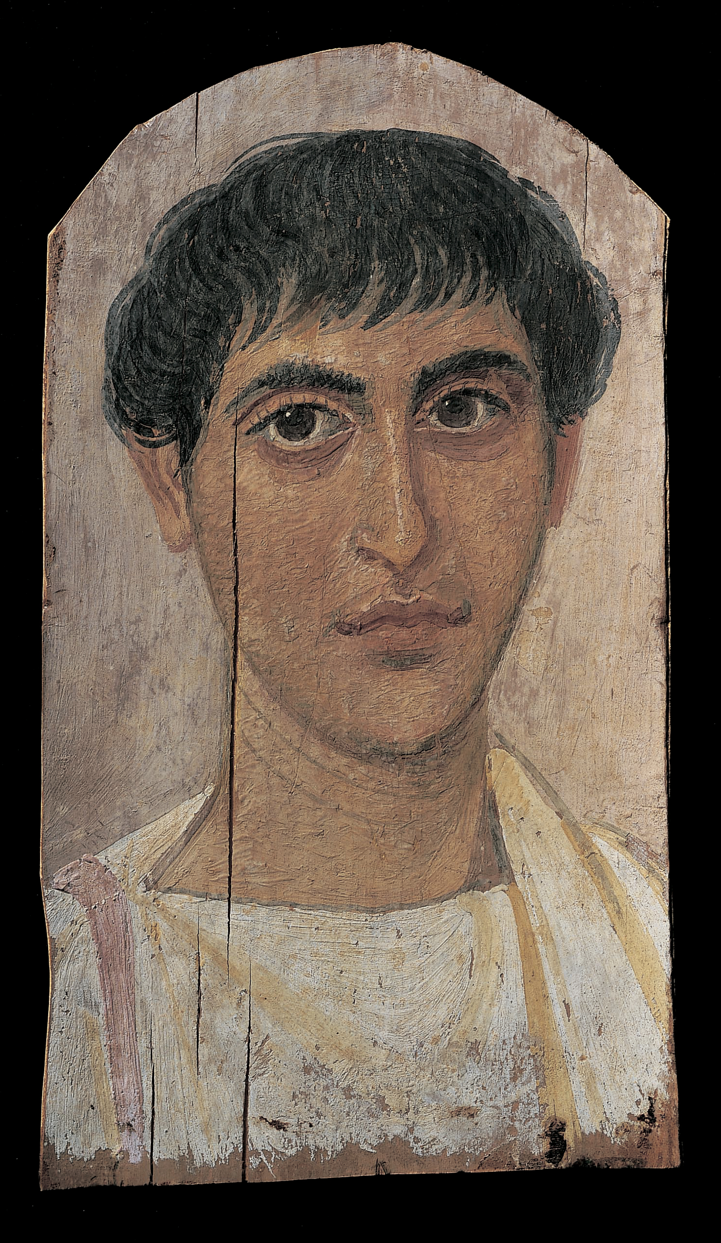
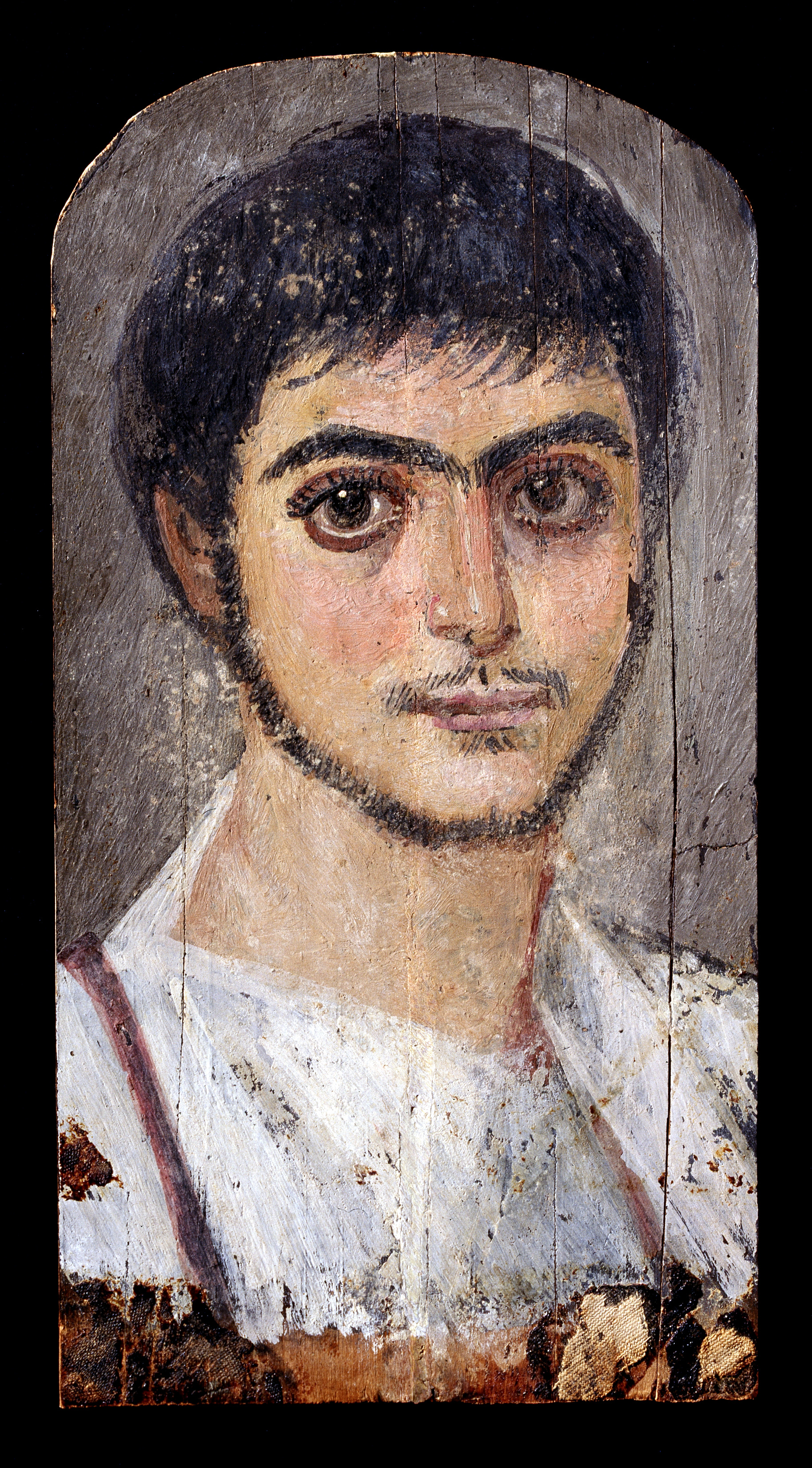
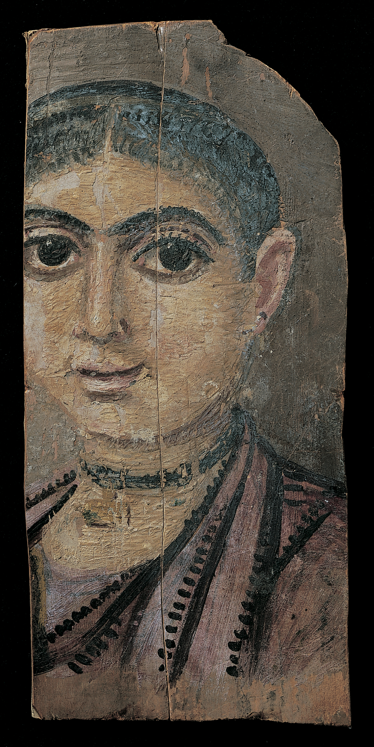
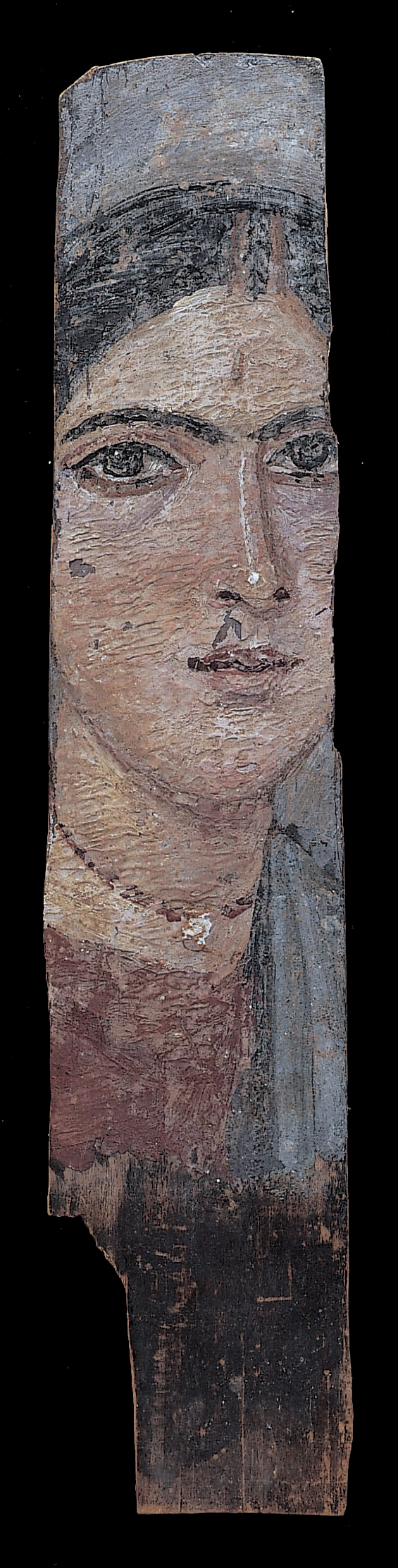
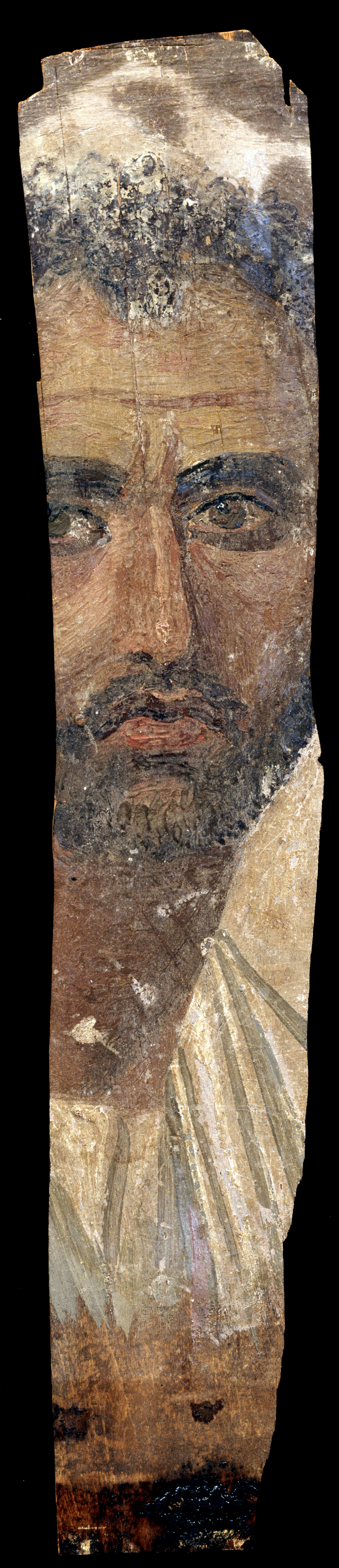
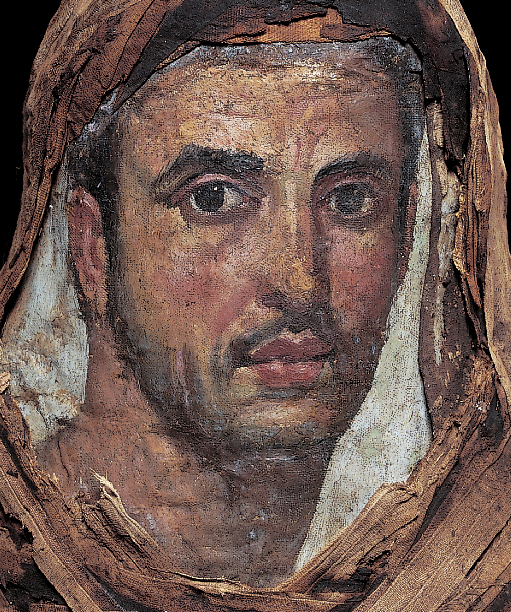
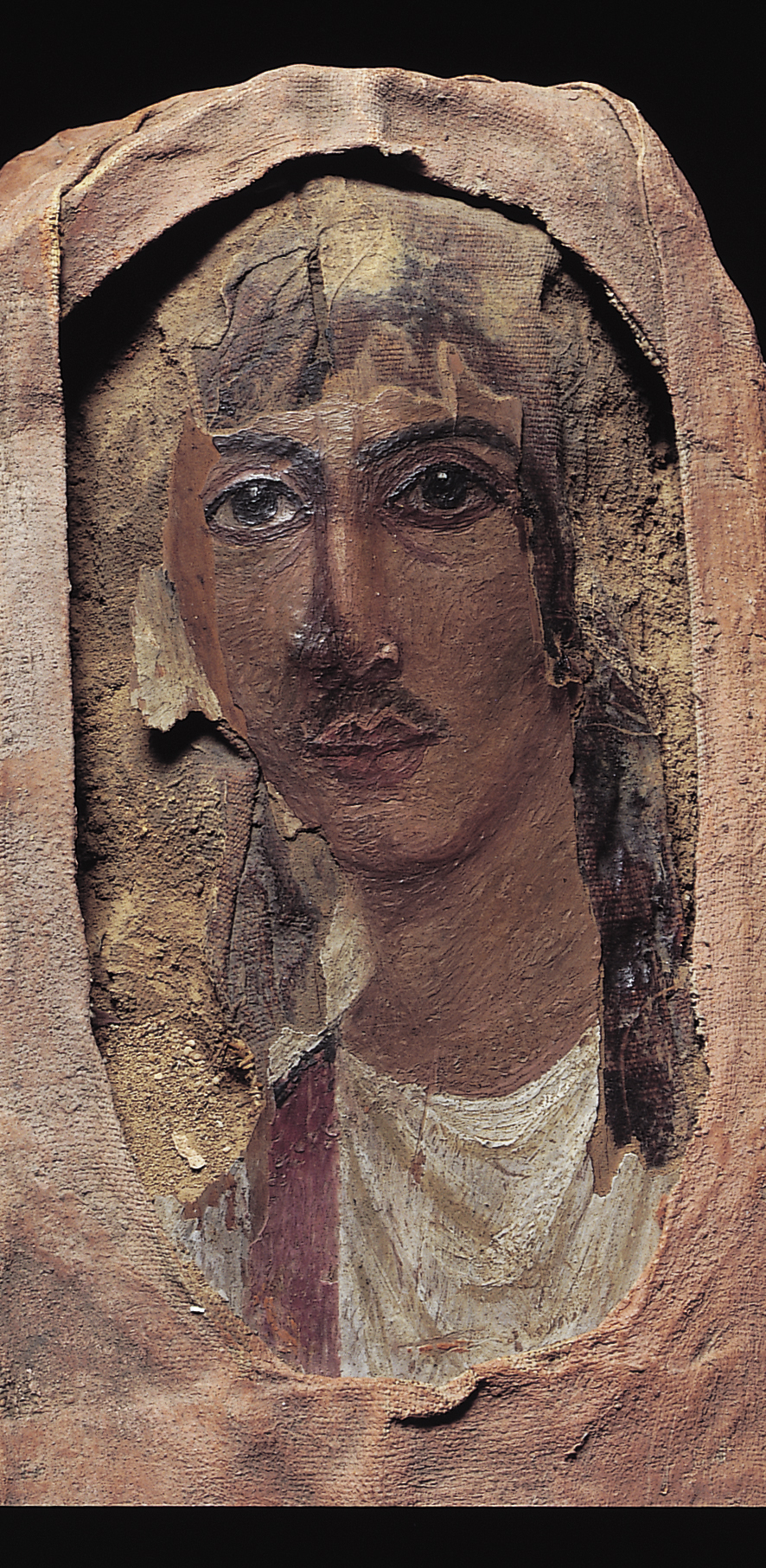
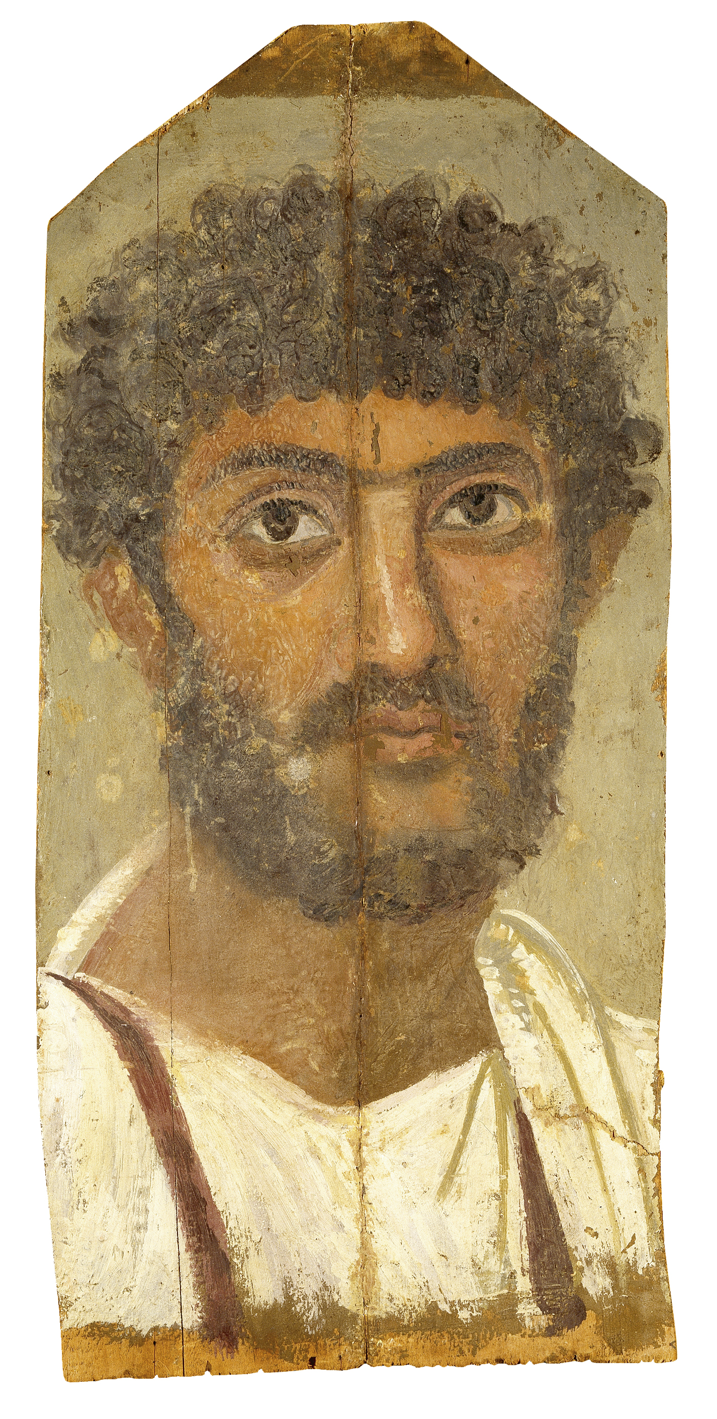
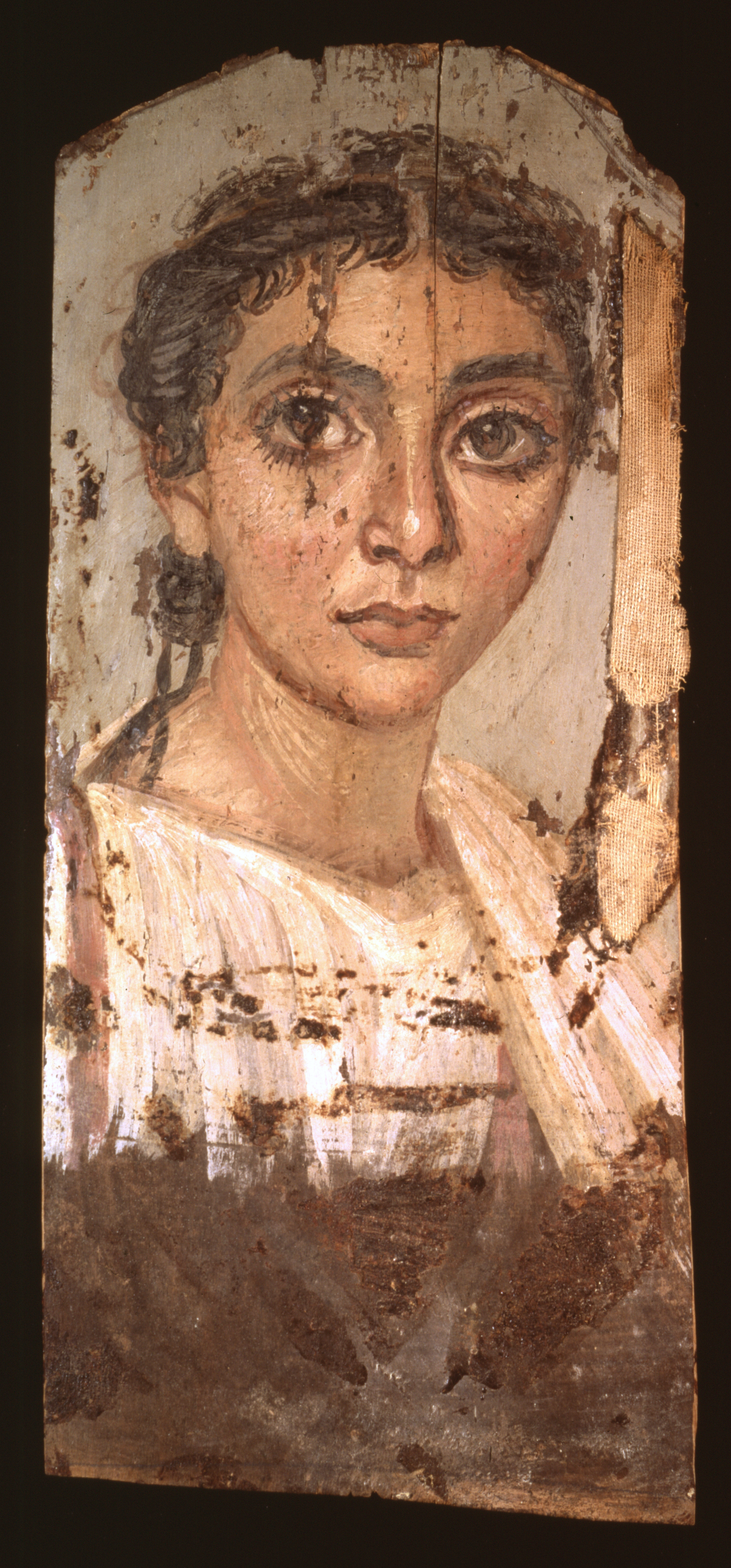
A study was designed to investigate the composition of beeswax and pigments from mummy portraits with the aim of improving our existing knowledge of the encaustic painting technique. This collaborative endeavor was made possible by the APPEAR project, which fosters interdisciplinary research between scientists and conservators and has resulted in new research yielding a better understanding of the inherent complexities of studying beeswax paint media. Colored paint samples were taken from nine encaustic mummy portraits (with different provenances) from two Danish museums, Ny Carlsberg Glyptotek (ÆIN) and the National Museum of Denmark (AS). Two portraits were excavated in Hawara by Sir W. Flinders Petrie, another five were bought from art dealer Theodor Graf’s collection and are suggested to come from er-Rubayat, and the provenience of the last two portraits has not yet been established but according to museum records they are assumed to also derive from er-Rubayat. Dating for the nine portraits varies; the oldest are assumed to be from 25–75 CE, the youngest 150–200 CE (fig. 9.1). The research presented here complements a previous study of binding media from the same portraits.1
| Collection | Ny Carlsberg Glyptotek | National Museum of Denmark | |||||||
|---|---|---|---|---|---|---|---|---|---|
| Provenience | Er-Rubayat (Graf collection) | Hawara (Petrie) | Presumable er-Rubayat | ||||||
| 680 | 681 | 682 | 683 | 684 | 1425 | 1426 | 3891 | 3892 | |
|
|
|
|
|
|
|
|
|
|
| Date | 100–125 CE | 125–150 CE | 140–160 CE | 125–150 CE | 140–200 CE | 25–75 CE | 100–125 CE | 150–200 CE | 150–200 CE |
| Flesh tone |
Lead white, iron oxides, carbon black,
kaolinite, goethite, minor red lead |
Iron oxides, lead white, kaolinite, carbon black, red lake |
Iron oxides, lead white, Egyptian blue, carbon black, quartz |
Iron oxides, lead white, carbon black, red lake, quartz |
Iron oxides, lead white, carbon black, kaolinite |
Iron oxides, lead white, Egyptian blue, carbon black, red lake, quartz |
Iron oxides, lead white, Egyptian blue, carbon black |
Iron oxides, lead pigment, carbon black, calcite, gypsum |
Egyptian blue, iron oxides, lead white, carbon black |
| Background | Lead white, iron oxides, carbon black |
Iron oxides, lead white, Egyptian blue, carbon black, quartz |
Iron oxides, lead white, carbon black, kaolinite |
Iron oxides, lead white, carbon black, kaolinite,
calcite |
Iron oxides, lead white, carbon black, |
Iron oxides, lead white, Egyptian blue, carbon black |
Iron oxides, lead white, Egyptian blue, carbon black |
Iron oxides, lead pigment, carbon black, calcite, gypsum |
Iron oxides, lead white, gypsum |
| Chiton |
Lead white, iron oxides, carbon black |
Iron oxides, lead white, kaolinite, carbon black |
Gypsum, red lake, bassanite, minor red lead, lead white |
Blue: Indigo, gypsum, bassanite, lead white Red: iron oxides, lead pigments, carbon black |
Iron oxides, lead white, carbon black, kaolinite, quartz | - | Lead white |
Iron oxides, lead pigment, carbon black, calcite, gypsum |
Iron oxides, lead white |
| Lips/red | Iron oxides, lead pigment, kaolinite |
Iron oxides, lead white, red lake (lips), calcite | Iron oxides, lead white | Red lead, lead white, iron oxides, kaolinite | Iron oxides, lead white, carbon black |
Vermilion, lead white, iron oxides, carbon black |
Hematite |
Iron oxides, lead pigment, carbon black, calcite, gypsum |
Iron oxides, lead white, carbon black |
| Hair/black |
Iron oxides, carbon black, lead white, red lake, kaolinite, quartz |
Carbon black, iron oxides, lead white, quartz | Carbon black, iron oxides, lead white, quartz | Carbon black, iron oxides, lead white, quartz | Iron oxides, lead white, carbon black, kaolinite | Carbon black, iron oxides, lead white | Carbon black, red lake, lead white, iron oxides, quartz |
Iron oxides, lead pigment, carbon black, calcite |
Iron oxides, lead white, gypsum, carbon black |
| Gold imitation | - | - | Goethite | Goethite | - | - | - | - | - |
| Clavus | Bassanite, red lake, lead white | Gypsum, red lake, bassanite, lead white | - | Red lead, red lake (minor), lead white, gypsum | Bassanite, gypsum, lead white, red lake | Lead white, red lake, iron oxides | Gypsum (?), lead white (minor), red lake, iron oxides (minor) | Lead white, iron oxides | Lead white, red lake |
| Ground | - | Carbon black, iron oxides | - | Iron oxides, carbon black | - | Calcite, gypsum, carbon black | - |
Iron oxides, gypsum (anhydrite), hematite |
Iron oxides |
The encaustic wax painting technique on some mummy portraits utilized heated and melted beeswax applied with small metal instruments leaving behind characteristically shaped and raised tool marks. Previous researchers have distinguished between encaustic technique and a beeswax technique executed in a painterly style that appeared more blended, with subtle variations in color and less impasto.2 This painterly beeswax technique has been described in the literature as “cold, modified, Punic or emulsified” and has caused misunderstanding of both the artist’s intent and technique. Pliny’s description of Punic wax has led to a belief among some scholars that the beeswax was (partially) saponified prior to its use in paints, allowing it to be applied cold, as an emulsion, and that various findings from analytical studies have been used to promote this idea.3 Still, the nomenclature and characterization of beeswax paint media is a source of contention and confusion.4,5 For example, the mummy portrait ÆIN 1425 was described as “wax used cold.”6
Typical beeswax is composed of 14% hydrocarbons, 35% palmitic wax esters, and 12% free fatty acids.7 Researchers have analyzed beeswax and paint samples from a small number of portraits executed in a painterly style (assumed to be modified or “used cold”) and concluded that a reduction in wax esters compared to hydrocarbons, the presence or absence of free fatty acids, or the detection of fatty acid metal soaps can be used to characterize “cold” applied wax or “modified beeswax.”8 These conclusions seem inaccurate, as researchers within the APPEAR project have shown through the scientific analysis of over thirty wax-paint portraits that fatty acid soaps from beeswax-paint portraits are independent of painterly style.9 Data also showed that they had similar beeswax profiles—the hydrocarbons had either evaporated or lesser amounts remained, fatty acid and wax ester content were consistent depending on the pigment, and they could contain significant amounts of palmitic acid lead soaps. The study showed the impact of pigments on the formation of fatty acid soaps in beeswax paint and how hydrolysis of wax esters results in increased amounts of palmitic acid soap.
To better understand why this occurs, a new study was designed to investigate whether lead white pigment impacts the formation of palmitic acid soaps. We hypothesized that lead white paint, a strong metal soap-forming pigment, would correlate to the presence of lead palmitate soaps in beeswax. The experimental methodology would quantify the amount of lead pigment and compare it to the palmitic acid metal soaps in beeswax paint. Fatty acid metal soaps have been used in beeswax paint to identify the cold wax technique, and the goal of this study was to provide a critical evaluation of this approach. Paint samples that varied in color, between light and dark, were tested for relative amounts of lead, wax esters, and palmitic acid soaps.
Method
Beeswax paint samples that were tested in this study were previously analyzed by gas chromatography/mass spectrometry (GC/MS) for palmitic acid and wax ester content. Details of the GC/MS results and methodology are described elsewhere.10,11 Briefly, the relative amounts of palmitic acid, wax esters, and other saturated and unsaturated monocarboxylic fatty acids in the samples were calculated from their chromatographic peak areas, expressed as percentages of the summed peak areas for all species measured. In this current study, the same samples were mounted and prepared as cross sections from the remaining material that was previously tested by GC/MS, and we focused on paint samples that varied in color between light and dark. Due to these inherent limitations, it was only possible to produce cross sections of both dark and light paint from three portraits. To enrich our data set and sample size, we tested variable colored paint samples from other portraits.
All cross sections were first photographed in visible light and with ultraviolet radiation and were subsequently examined by scanning electron microscopy–energy dispersive X-ray spectroscopy (SEM-EDX).12 The samples’ surfaces were scanned and the amount of lead in the paint sample13 was calculated as a percent value based on the sum of a variable pigment mixture depending on the portrait, color, and technique. The lead percentage depends on the sample location and, therefore, the amount of lead pigment found in the cross section. Lead white is the most common pigment used in beeswax-based portraits and will readily form soaps with palmitic acid and other fatty acids.
Results and Discussion
As expected, GC/MS results from wax paint samples showed that lighter tones (white or flesh colored) contained more palmitic acid soap compared to wax esters (fig. 9.2). Conversely, the darker paint samples contained lower percentages of palmitic acid soap. For example, two paints were selected from the same portrait—ÆIN 682: black contains 8% palmitic acid soap and 54% wax esters and flesh tone areas contain 41% palmitic acid soap and 18% wax esters. Two exceptions in this study were white areas on 3892 (AS) and the flesh tone areas of ÆIN 681, as they did not contain the larger percentage of palmitic acid soaps. Most lighter-colored paints, however, such as the flesh tones on ÆIN 682, did contain significantly more palmitic acid soap relative to wax esters. We propose that the relatively large amounts of palmitic acid soap in lighter-colored paints are due to the presence of lead white.
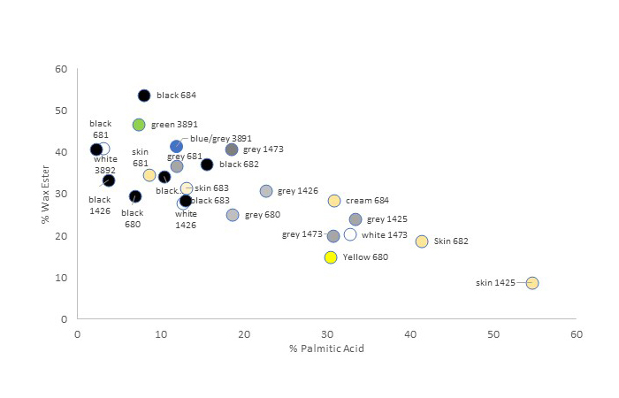
Beeswax is composed of wax esters, which contain palmitic acid (fatty acid) and a long-chain alcohol. Hydrolysis of the wax ester produces free palmitic acid and alcohols, and if lead pigment is present, lead palmitate (fatty acid soap) will form. Fatty acid soap formation is a well-studied phenomenon in oil paintings and primarily occurs with lead and zinc. However, magnesium, aluminum, calcium, and copper pigments also form metal complexes with free fatty acids.14,15 Lead soaps may disrupt the paint matrix and form protrusions but can also stabilize the fatty acids within a paint matrix and speed up drying time. Without forming metal soaps, free fatty acids will eventually migrate out of the paint, evaporate, or leave a white efflorescence or ghost images if under glass.16 Over time, and depending on the pigment present, the chemical profile of old beeswax paint is modified when compared to fresh beeswax. Lead white paint will contain significantly more palmitic acid soap, while paint samples without lead white (or metal-forming pigments) do not form lead soaps, resulting in the evaporation of fatty acids from the paint matrix.
SEM-EDX and GC/MS results for percentages of lead and palmitic acid are shown in figure 9.3, where results for each sample are described as a color with its portrait number. The percentage of lead in the paint samples was calculated based on the estimated content and sum of a variable mixture of pigments using SEM-EDX. Therefore, both values may be affected by the inhomogeneity of the paint sample and application methodology employed. Preliminary results indicate that lighter-colored samples contain significantly higher percentages of lead and palmitic acid soap, while the opposite is true for darker-colored samples. For example, the flesh tones in the ÆIN 682 portrait contain approximately 15% lead and 43% palmitic acid, while ÆIN 682 black contains 3% lead and 16% palmitic acid.
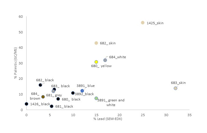
It should be noted that several samples were outliers and did not follow the trend of increased lead and palmitic acid content, such as the flesh tones on ÆIN 683. The variability of our data could be due to several reasons. First, the inhomogeneity of the pigment particles was evident in the area/surface of the SEM-EDX scan. When polishing the cross sections, the pigment particle size was highly variable and the size of the particles at the surface of the cross section changed with subsequent polishing. This change could impact the results of the SEM-EDX scan on a given cross section and may indicate an area of inhomogeneity that is not representative of the lead pigment content. Second, a small cross section sample does not always represent the lead content found in larger areas of the portrait. Third, it is well known in oil-paint studies that, due to the soaps’ ability and tendency to migrate throughout the paint layers, lead soaps form concentrated inclusions in areas within the paint matrix.17 This would result in variable amounts of palmitic acid detected in a small paint sample compared to an overall average in a larger paint area.
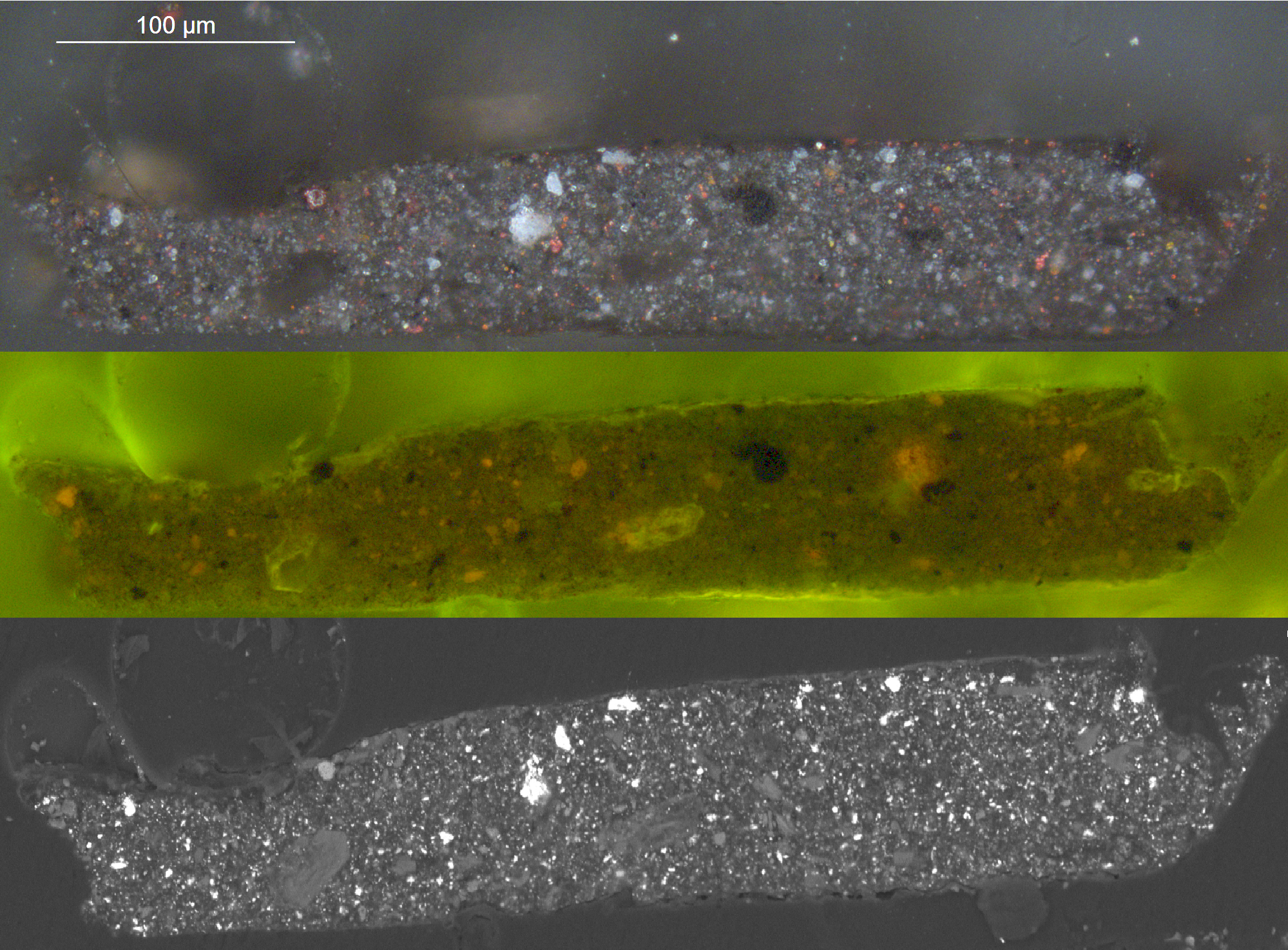
The following pigments were identified in the seven dark paint samples: carbon black, red/brown iron oxides, and, in one example, red lake. The SEM-EDX cross section scans provided information on the pigments used in the specific paint layers studied. Figure 9.4 shows an example of a cross section from a dark color, ÆIN 1426, and illustrates the complexity of pigments found in dark or black areas. The visible light image shows how the paint is composed of black, white, brown, red, and yellow pigments. There is a surprisingly high lead white content (seen as white particles in the SEM-EDX image) in many of the dark/black paint layers. The cross section also shows the presence of red lake pigment, a surprising addition to black paint. The red lake pigments can be observed as bright orange particles with or without ultraviolet radiation.
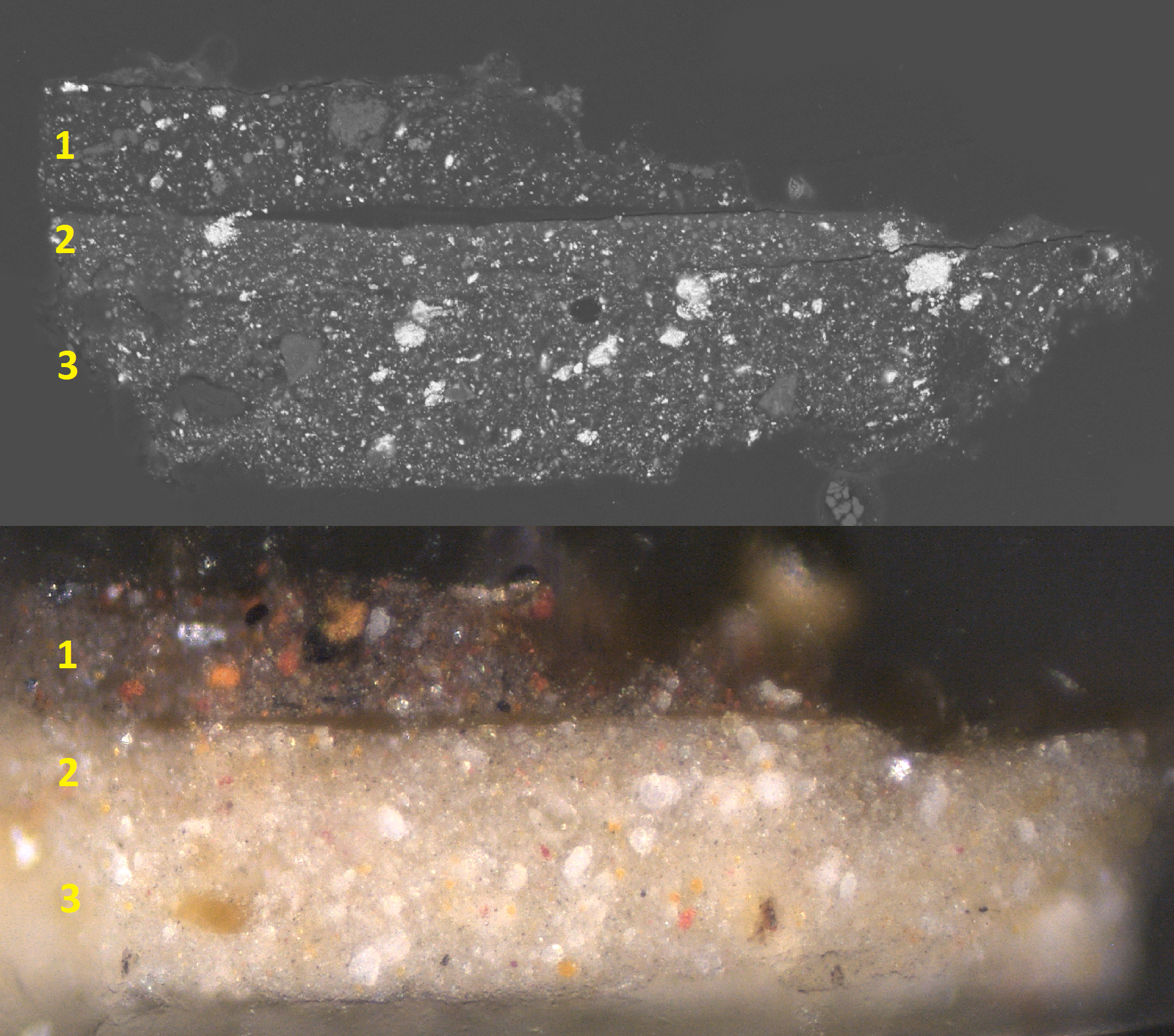
The cross sections also gave interesting information on the stratigraphy of the wax paint, though most samples were diminutive and thus did not contain all the layers in the paint structure. However, a cross section shows the stratigraphy of black hair (layer no. 3) on top of two layers (nos. 1 and 2) of light gray background color from ÆIN 681 (fig. 9.5). The visible light image shows how both the light and dark colors consist of red, yellow, black, and white pigments, and in most cases yellow iron oxide as well. Calcite and quartz were present in all dark-colored samples as well as lead white in surprisingly significant amounts when compared to the dark sections of the paint. It is noteworthy that we did not detect the red pigment minium in the darker areas, however, lead white was ubiquitous in all the samples analyzed and is the only pigment present in the paint samples capable of forming lead soaps.
Lead white, calcite, and quartz mixed with various hues of red and yellow iron oxides were observed in all six light-colored samples analyzed. The addition of Egyptian blue was also identified in two samples taken from light-colored flesh tones (see fig. 9.1). The pigments identified in this study of mummy portraits painted with beeswax-based paint revealed that the painters had an economical yet precise and conservative palette of pigments, and the results corroborate what other studies have so far reported.
Conclusion
The SEM-EDX results revealed that both dark and light beeswax samples taken from nine beeswax-based mummy portraits contained variable amounts of lead white. Carbon black, with the addition of red/brown iron oxides and one example of red lake, was identified in the seven dark paint samples. Lead white, calcite, and quartz mixed with various hues of red and yellow iron oxides were observed in light-colored paints.
Through a fortuitous collaboration, evidence is presented here that allows for a better understanding of how lead soaps form in ancient beeswax and why its presence should not be used as an indication of cold applied or Punic wax. GC/MS and SEM-EDX results were compared and preliminary results show that lead is positively correlated with the formation of palmitic acid soaps. This provides further evidence that the soaps present in beeswax paint can be attributed to lead metal complexes. Therefore, differences in beeswax chemical profiles should not be used to indicate a purposeful modification of the beeswax during the preparation of the beeswax paint. Rather, the beeswax chemical profiles are different from one another because of lead content, evaporation, pigments, wax ester hydrolysis, and subsequent soap formation. The hydrolysis of wax esters occurs over time and, when palmitic acid is released, it forms a palmitic acid lead soap. This explains the variation we see in the wax ester profile, and it is hoped that these results will inspire discussion and offer a clearer understanding of the ancient beeswax painting technique.
Acknowledgments
The authors are grateful to several individuals for their contribution to this study: Julie Jaeger, teaching associate professor, Royal Danish Academy for Fine Arts; Richard Newman, head of scientific research, Museum of Fine Arts, Boston; and Michelle Taube, conservation scientist, the National Museum of Denmark.
The authors would also like to thank the following entities for their support: the Carlsberg Foundation and the Getty Conservation Institute; the staff at the National Museum of Denmark and the Ny Carlsberg Glyptotek; the Molab team, led by Costanza Miliani; the APPEAR team and Marie Svoboda; and the Center for Art Technological Studies, National Gallery of Denmark.
Notes
-
Spaabæk, Lin Rosa, and Joy Mazurek. 2020. “Binding Media and Coatings: Mummy Portraits in the National Museum of Denmark and the Ny Carlsberg Glyptotek.” In Svoboda and Cartwright 2020.. ↩︎
-
Ramer, Brian. 1979. “The Technology, Examination and Conservation of the Fayum Portraits in the Petrie Museum.” Studies in Conservation 24 (1): 1–13.. ↩︎
-
Kühn, Hermann. 1960. “Detection and Identification of Waxes, Including Punic Wax by Infrared Spectrography.” Studies in Conservation 5 (2): 71–81. https://doi.org/10.2307/1504955.. ↩︎
-
Sutherland, Ken, Rachel C. Sabino, and Federica Pozzi. 2020. “Challenges in the Characterization and Categorization of Binding Media in Mummy Portraits.” In Svoboda and Cartwright 2020.. ↩︎
-
Stacey, Rebecca J. 2011. “The Composition of Some Roman Medicines: Evidence for Pliny’s Punic Wax?” Analytical and Bioanalytical Chemistry 401 (6): 1749–59.. ↩︎
-
Doxiadis, Euphrosyne. 1995. The Mysterious Fayum Portraits: Faces from Ancient Egypt. Thames & Hudson.. ↩︎
-
Jiménez, Juan J., José L. Bernal, S. Aumente, María Jesús del Nozal, María Teresa Martín, and José Bernal Jr. 2004. “Quality Assurance of Commercial Beeswax. Part I. Gas Chromatography–Electron Impact Ionization Mass Spectrometry of Hydrocarbons and Monoesters.” Journal of Chromatography A 1024 (1–2): 147–54.. ↩︎
-
White, Raymond. 1978. “The Application of Gas-Chromatography to the Identification of Waxes.” Studies in Conservation 23 (2): 57–68. https://doi.org/10.2307/1505796.. ↩︎
-
Mazurek, Joy, Marie Svoboda, and Michael Schilling. 2019. “GC/MS Characterization of Beeswax, Protein, Gum, Resin, and Oil in Romano-Egyptian Paintings.” Heritage 2 (3): 1960–85. https://doi.org/10.3390/heritage2030119.. ↩︎
-
Spaabæk, Lin Rosa, and Joy Mazurek. 2020. “Binding Media and Coatings: Mummy Portraits in the National Museum of Denmark and the Ny Carlsberg Glyptotek.” In Svoboda and Cartwright 2020.. ↩︎
-
Mazurek, Joy, Marie Svoboda, and Michael Schilling. 2019. “GC/MS Characterization of Beeswax, Protein, Gum, Resin, and Oil in Romano-Egyptian Paintings.” Heritage 2 (3): 1960–85. https://doi.org/10.3390/heritage2030119.. ↩︎
-
S-3400N scanning electron microscope from Hitachi combined with a Bruker XFlash 6I30. ↩︎
-
The pigment identifications were done by various methods: internal reports by MOLAB, led by C. Miliani (FTIR, XRF, UV-vis, SEM-EDX, Raman) (see also Miliani, Costanza, Alessia Daveri, Lin R. Spaabæk, Aldo Romani, Valentina Manuali, Antonio Sgamellotti, and Brunetto G. Brunetti. 2010. “Bleaching of Red Lake Pigments in Encaustic Mummy Portraits.” Applied Physics A 100 (3): 703–11. https://doi.org/10.1007/s00339-010-5748-3.); internal report by the Danish National Museums research department, by chemist M. Taube (XRF analyses) and M. C. Christensen (amino acid and FTIR analyses); internal reports by R. Newman, MFA, Boston (FTIR, Raman, SEM); internal report by D. Buti, CATS, National Gallery of Denmark (XRF); internal report by Julie Jaeger, teaching associate professor, KADK (SEM-EDX); Nini A. Reeler, chemist, University of Copenhagen (Raman) (see also Reeler, Nini Elisabeth Abildgaard, Ole Faurskov Nielsen, Lin Spaabæk, Mogens Jørgensen, and Henrik Grum Kjaergaard. 2013. “Pigments and Binding Material in Fayum Mummy Portraits Determined by NIR-FT-Raman Microscopy.” Asian Chemistry Letters 17: 1–12.). The portraits have also been studied by various analytical photographic methods: visible-induced infrared luminescence (VIL), X-radiography, and ultraviolet radiation. Figure 9.1 shows a schematic overview of pigment identification. ↩︎
-
van den Berg, Jorrit D. J., Klaus Jan van den Berg, and Jaap J. Boon. 1999. “Chemical Changes in Curing and Ageing Oil Paints.” In ICOM Committee for Conservation 12th Triennial Meeting Lyon 29 August–3 September 1999. James & James.. ↩︎
-
Casadio, Francesca, Katrien Keune, Petria Noble, Annelies Van Loon, Ella Hendriks, Silvia A. Centeno, and Gillian Osmond. 2019. Metal Soaps in Art. Springer International.. ↩︎
-
Schilling, Michael R., David M. Carson, and Herant P. Khanjian. 1998. “Evaporation of Fatty Acids and the Formation of Ghost Images by Framed Oil Paintings.” WAAC Newsletter 21 (1). https://cool.culturalheritage.org/waac/wn/wn21/wn21-1/wn21-106.html.. ↩︎
-
Plater, M. John, Ben De Silva, Thomas Gelbrich, Michael B. Hursthouse, Catherine L. Higgitt, and David R. Saunders, 2003. “The Characterisation of Lead Fatty Acid Soaps in ‘Protrusions’ in Aged Traditional Oil Paint.” Polyhedron 22 (24): 3171–79. https://doi.org/10.1016/S0277-5387(03)00461-3.. ↩︎