The core of the Egyptian collection of the National Archaeological Museum (NAM) in Athens consists of significant donations from two Greek expatriates from Egypt: Ioannis Dimitriou, in 1880, and Alexandros Rostovich, in 1904. The collection was further enriched with donations from the Egyptian government, in 1893, and the Greek Archaeological Society, in 1894. Among the exhibited artifacts of the Egyptian collection are five funerary portraits. Two are on fabric backings; the other three are polychromed wooden panels. The portraits lack archaeological contexts. The initial discovery sites of these works are not known and, unfortunately, are not preserved along with associated burial finds. Moreover, no further scientific examination of their state of preservation, of previous interventions, and of the manufacturing methods and materials has ever been carried out. Therefore, it soon became obvious to the NAM that a more in-depth investigation of the works was both needed and urgent; the museum’s participation in the APPEAR project brought about the opportunity for further study.
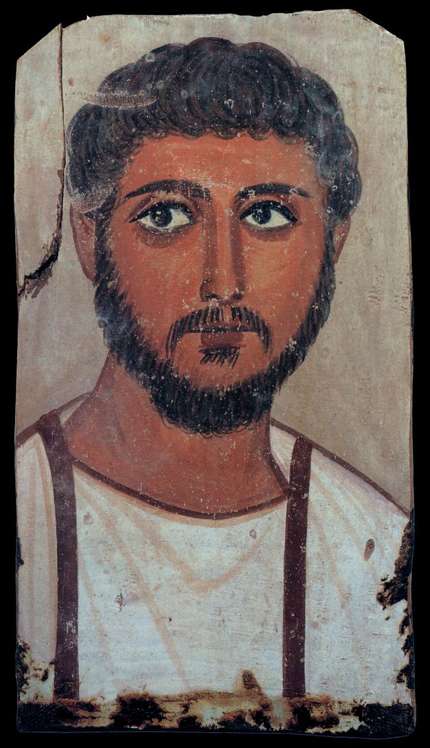
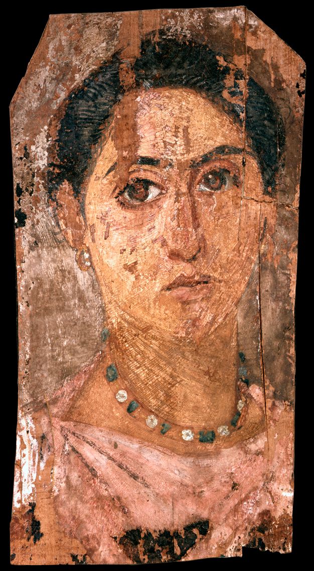
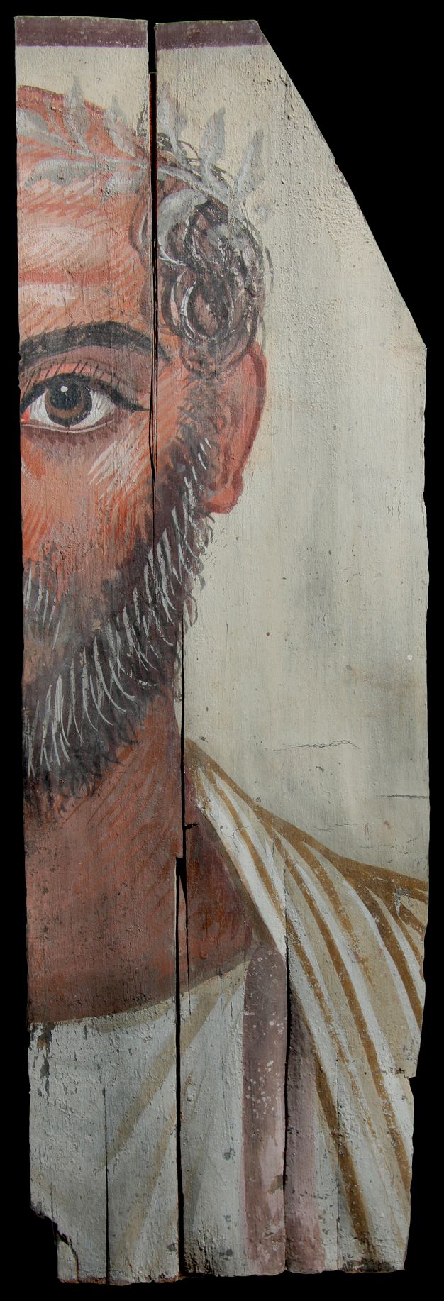
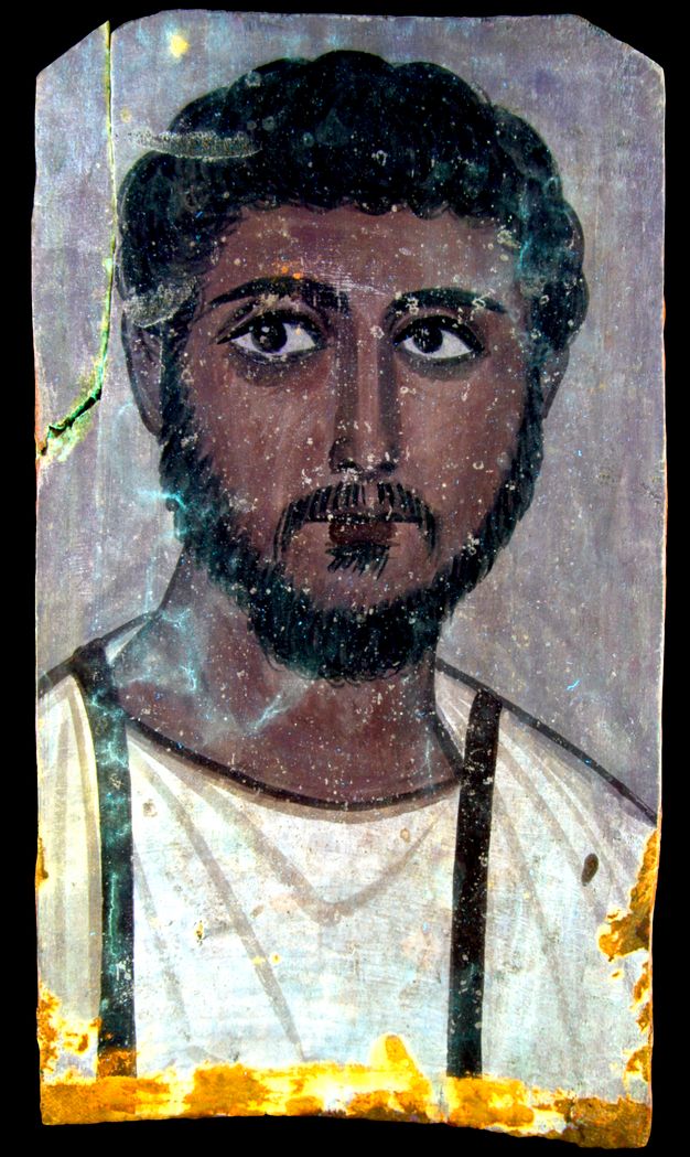
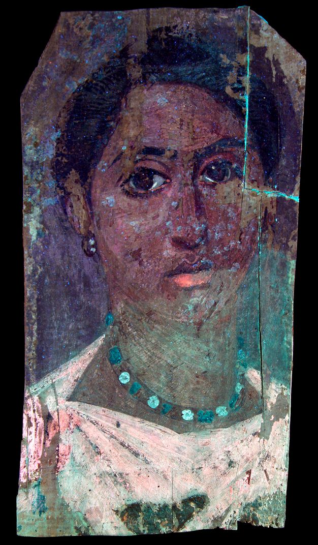
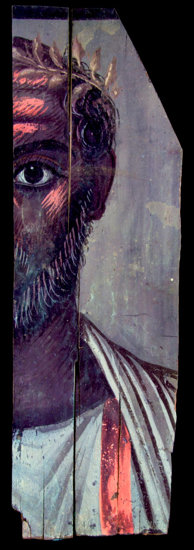
For the museum’s initial participation in the APPEAR project the three portraits on wooden panels were chosen. The first depicts a young man (inv. no. 1627); the second, an older man (inv. no. 1629); and the third, a woman (inv. no. 1628; figs. 5.1–5.3). Although no analysis of the binding media was performed, the two male portraits appear to be painted in tempera and the female portrait in encaustic.1 Macroscopically, the two male portraits appear very similar in terms of manufacture and style, while the female portrait exhibits more elaborate workmanship. Previous conservation and restoration interventions are obvious in the portraits of the young man and the woman: split wood fragments have been adhered in both.
Using instruments available at the NAM, the present study involved multiple approaches with consecutive steps. First and foremost, we employed multiband imaging (MBI) and reflectance transformation imaging (RTI) to document in detail the works’ painted surface. In the next step, X-radiography investigated both surface characteristics and items of initial manufacture or later intervention. Portable X-ray fluorescence (pXRF) spectroscopy followed for the elemental analysis of the painted surface. We decided to examine many spots across the painted surface to acquire information on a larger area, which produced results similar to those obtained by the XRF mapping. Finally, a digital microscope, used in a handheld “scanning mode,” helped us to interpret the results obtained by some of the other aforementioned methods.
Imaging
For imaging purposes, we used MBI and X-ray radiography. RTI is a very powerful tool that, in short, combines raking light photography with computational algorithms to digitally examine surface details. For the portraits, RTI shed light on the state of preservation of the works’ wooden supports and paint surface as well as on the methods of color application.
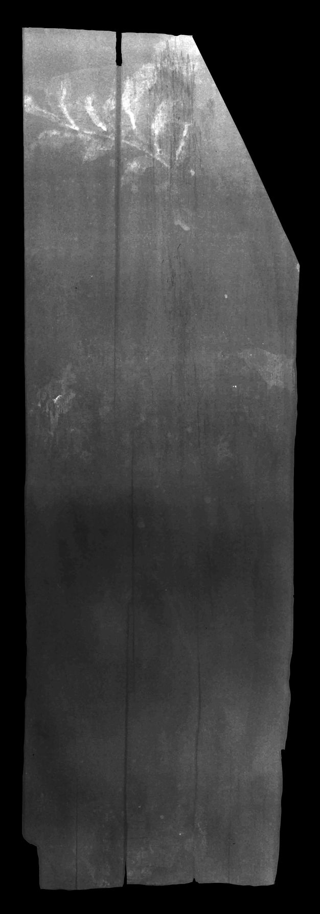
RTI revealed that significant warping along the grain of the wooden support is present on the male portraits; also, cracks appear both on the supports and within the paint layers. Cracking is an issue on the female portrait panel too; however, in this case the severity of the compromised paint layer has resulted in a significant loss of paint. RTI emphasized the highly textured impasto layer on the female portrait, especially on the skin, facial features, and jewelry (fig. 5.4). On the two male portraits, in contrast, because all paint layers had been applied very thinly, all relief was limited to the wooden support.
MBI carried out on all three portraits consists of images captured in the visible spectrum; fluorescence in the visible region, generated by ultraviolet-induced visible fluorescence (UVF); and reflectance in the near infrared region, produced by infrared reflectography (IRR) and visible-induced luminescence (VIL). Figure 5.5 summarizes the imaging methodology used.
| Technique | Camera | Light source | Filters |
|---|---|---|---|
| VIS | CANON EOS 700D | Daylight | BRAUN Blueline UV Lens Protection |
| UVF | CANON EOS 700D | SYLVANIA Blacklight-Blue F18W/BLB-T8 Tube (315nm–400nm) | BRAUN Blueline UV Lens Protection |
| IRR | CANON EOS 20D Modified | Halogen Flood light IP44 400W | LVSHI IR850nm |
| VIL | CANON EOS 20D Modified | LUMAX Plati LFL107 LED Flood light 4500lm, 6000K | LVSHI IR850nm |
Fluorescence in the visible region (figs. 5.6–5.8) revealed undocumented previous interventions, some of which had never been observed before. The apparent assemblages of the separated fragments in the portraits of the young man and the woman were executed with probably a cellulose nitrate adhesive.2 For the paint surface’s consolidation, which for the most part had gone undetected, UVF photography proved immensely valuable; it showed both the extent of the treatment and the various materials used to carry it out—most likely at different times. The two male portraits were locally treated with what appears to be, based on its characteristic fluorescence, a polyvinyl acetate (PVAc) dispersion, which was likely applied in the same manner to the female portrait as well.3 However, the latter revealed at its lower half extensive consolidation with a different material, demonstrating the greenish fluorescence of a plant-based resin, possibly dammar gum or mastic.4 The use of a natural resin for consolidation purposes suggests treatment at an earlier date than that of the PVAc application. Additionally, two small areas above the proper right (PR) eyebrow of the young man were treated with what appears to be shellac; however, because they are only two very small spots in an area that does not seem to have ever needed consolidation, we cannot rule out the possibility of accidental contamination when the portrait was kept in a conservation laboratory—or rather, a “mending workshop”—of that period. UVF also exposed the use of an organic lake, probably madder, on the facial features and garments of the older man (forehead, cheekbone highlights, clavus) and the woman (lips, eyes, tunic).
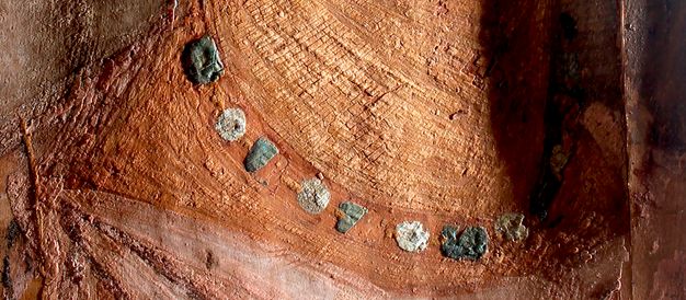
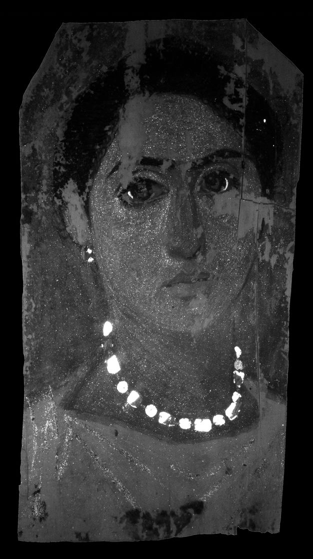
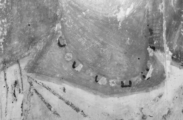
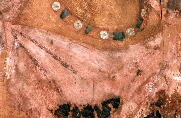
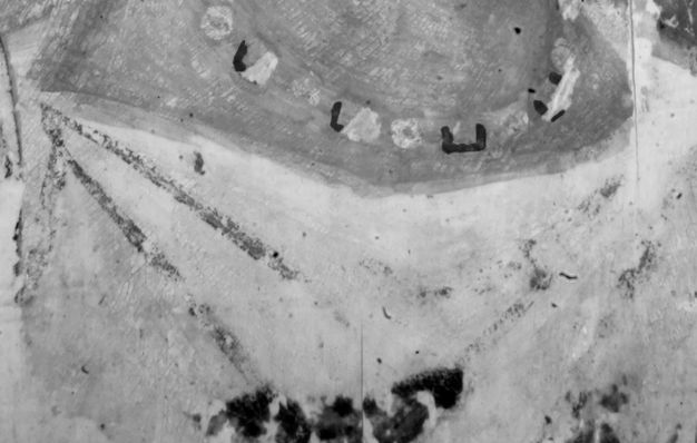
The VIL technique, employed for every portrait, produced positive results only for that of the female (see figs. 5.1 and 5.9). The spatial distribution of Egyptian blue on this work—apart from the obvious areas appearing in the visible spectrum with a green-blue hue—was observed in more unexpected areas: the white beads of the jewelry, the skin, and parts of the tunic and eyes. A point of interest is the fact that the luminescence of Egyptian blue appears equally bright among areas that are presented in totally different hues, as if the pigment application is of equal proportion. This effect is notably manifested in the green-blue and the white beads of the necklace and earring.
A reflectance response in the IR region suggests the addition of an organic black colorant for the darker areas such as the hair, eyebrows, and outlines for all three portraits. IRR also revealed the presence of underdrawings on the female portrait (fig. 5.10). A distinct outline, a half rectangle in shape, guides the placement of each of the green-blue beads of the necklace.
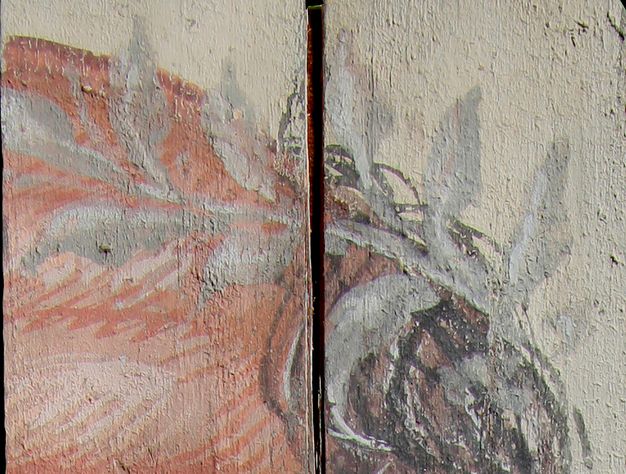
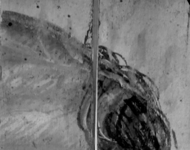
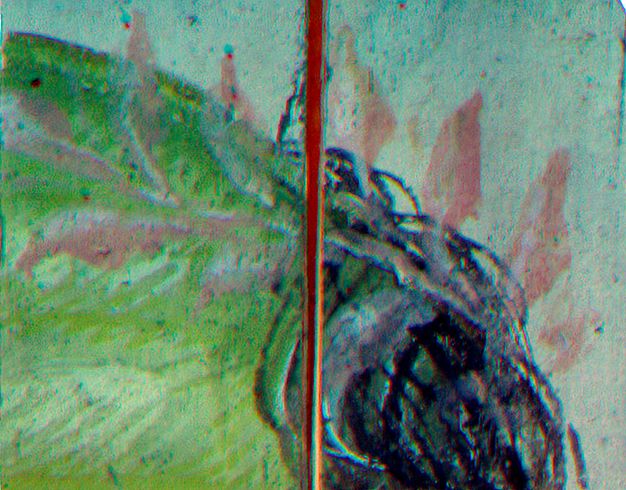
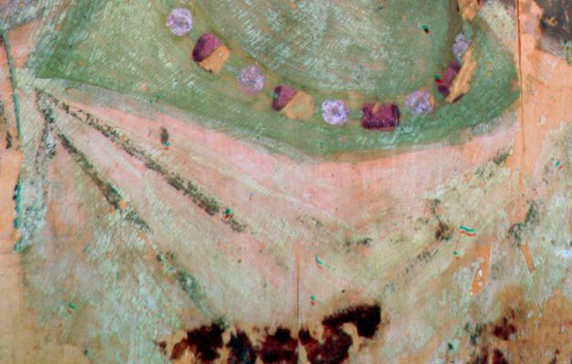
We also digitally combined the IRR and VIS images to create infrared-reflected false color (IRRFC) images.5 In the case of the older man’s wreath, the IRRFC image revealed a hue, mainly in the shading of the leaves, consistent with the response of Egyptian blue, indigo, and some green earths (figs. 5.11–5.13).6 In the case of the woman’s necklace and garment, the IRRFC image indicated that the green-blue beads and a few of the central folds corroborate the use of Egyptian blue—as seen in the VIL image. A wash of green earth or indigo is likely to have been added (figs. 5.14–5.16).7
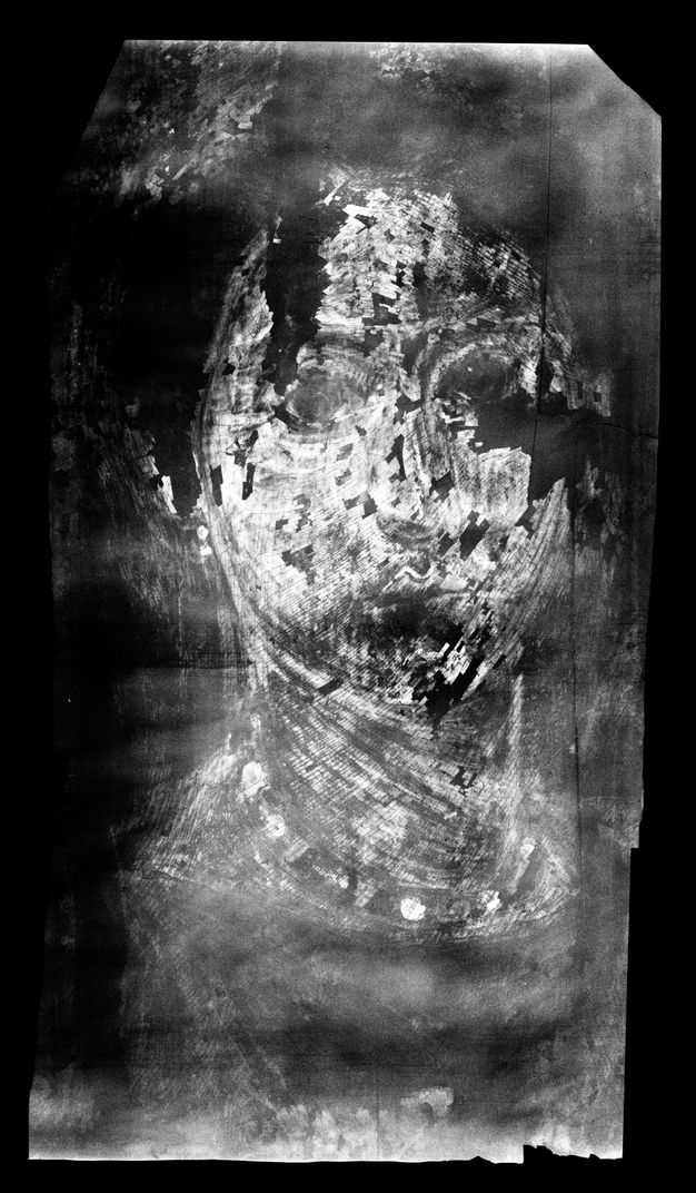
X-radiography was carried out using the in-house X-ray tube even though the equipment’s setup is intended for industrial applications. The resultant images (figs. 5.17 and 5.18) indicate the application of a lead-based pigment, most probably lead white, for most of the female portrait and for the older man’s wreath.
Chemical Analysis
Although imaging is a powerful tool for examining artifacts, it is regarded as a preliminary step in a scientific study; in-depth analysis is needed to obtain more precise results. For the portraits’ elemental assessment, we employed readily available instruments at the NAM. The portable TRACER 5i XRF spectrometer by Bruker, equipped with a live-view camera and an 8-millimeter collimator, was used and run at two of the manufacturer’s built-in applications—that is, Alloys 2 and MuddrockAir—so as to capture all possible chemical elements present on the paint surface. We also decided to examine between thirty-three and forty-five of the XRF targets’ locations on each portrait to acquire information from a larger surface area and to produce results that yield a map of the elements present. In addition to this mapping attempt, we separated the paint surfaces into groups according to their hue and uniformity during data capture. The mean, median, maximum, and minimum values of the elements were calculated for different areas—the background, hair, skin, etc.—of the three portraits. Figure 5.19 summarizes the portraits’ chemical elements according to their mean values.
| Portrait and Number of Targets | Group Areas | Major Elements | Minor Elements | Trace Elements |
|---|---|---|---|---|
|
Fig. 5.1 33 targets 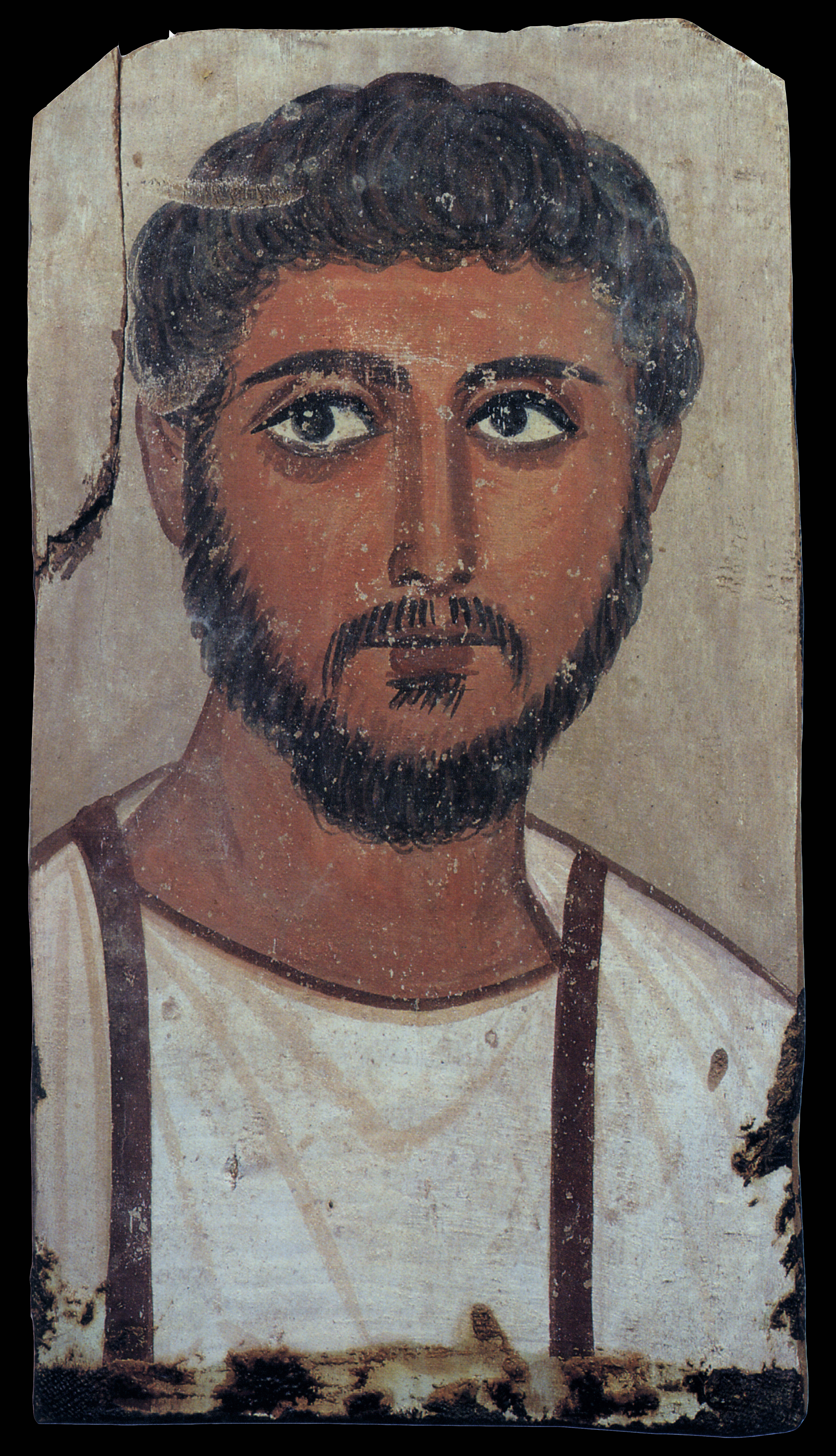
|
Background | Ca, S | Si, Al, | Fe, Mn, K, P |
| Hair | Ca, Fe, Si, S | Al, Mg | Mn, P | |
| Skin | Ca, S | Fe, Al, Si, Mg | Mn, K, P | |
| Lips | Ca, S, Fe, Si, Mg | Al | Mn, P | |
| Tunic | Ca, S | Fe, P, Si, Mg | ||
| Clavi | Fe, Ca, Si | As, S, Al, Mg | Mn, K, P | |
|
Fig. 5.2 45 targets 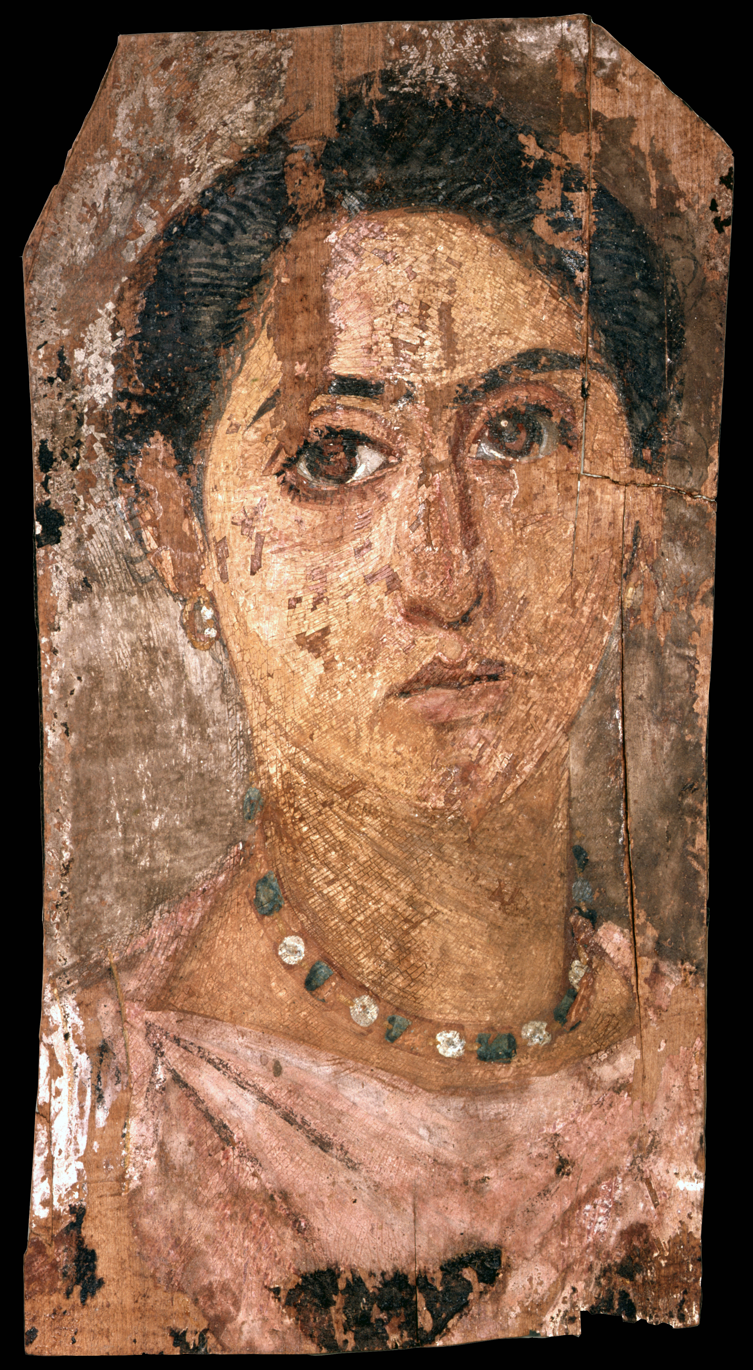
|
Background | Pb, Ca, S, Si | As, Fe, Mg | Cu, Mn, K, P, Al |
| Hair | Pb, Fe, Ca, S, Si | As, Mg | Cu, Mn, K, P, Al | |
| Eyebrows | Pb, As, Fe, S | Ca, Si, Mg | Cu, Mn, K, P | |
| Skin | Pb, As, Fe, S | Hg, Ca, Si, Al, Mg | Cu, Mn, K, P | |
| Lips | Ca, S, Si | Pb, As, Fe, Al, Mg | Cu, Mn, K, P | |
| Tunic | Ca, S, Al | Pb, Si, Mg | As, Cu, Fe, Mn, K, P | |
| Green beads | Pb, Cu, Fe, Ca, Si, Hg | As, S, Al, Mg | Mn, K, P | |
| White beads | Pb, As, S, Si | Cu, Fe, Ca, Mg | K, P, Al | |
|
Fig. 5.3 35 targets 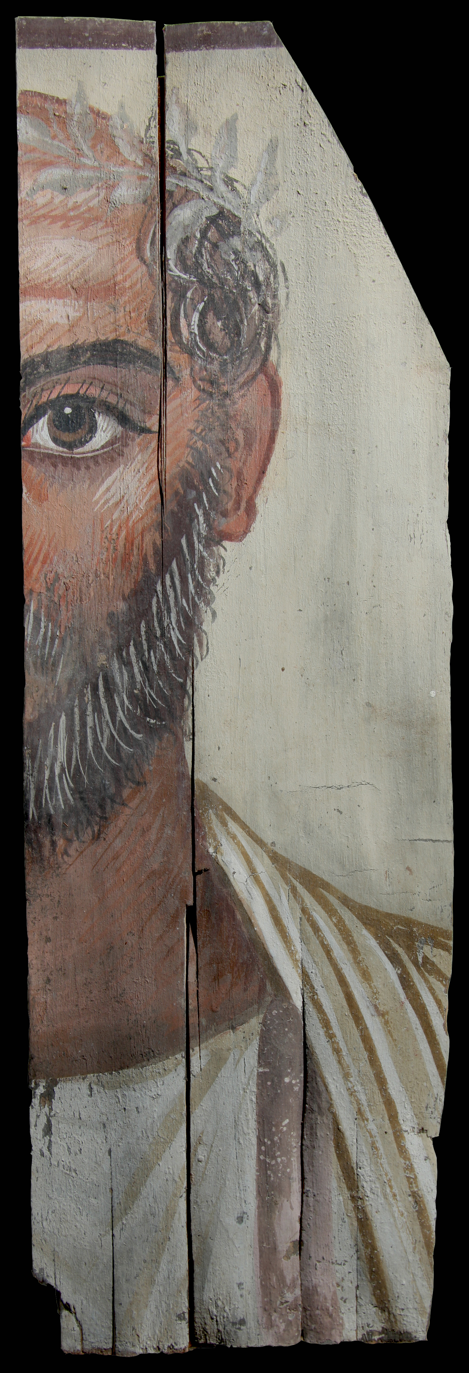
|
Background | S, Al | Pb, As, Fe, Ca, Ca, Si, K, Mg | Cu, Mn, P |
| Hair | Fe, S, Al | Ca, Si, Mg | Pb, As, Cu, Mn, K, P | |
| Skin | S, Al | Fe, Ca, Si | Pb, As, Cu, K, P, Mg | |
| Wreath | Pb, As, S | Ca, Cl, Si, Mg | Cu, Fe, Mn, K, P, Al | |
| Tunic | S, Al | Ca, Si | Pb, As, Cu, Fe, Mn, K, P, Mg | |
| Clavus | Ca, S, Si, Al | Pb, Fe, Mg | As, Cu, Mn, K, P |
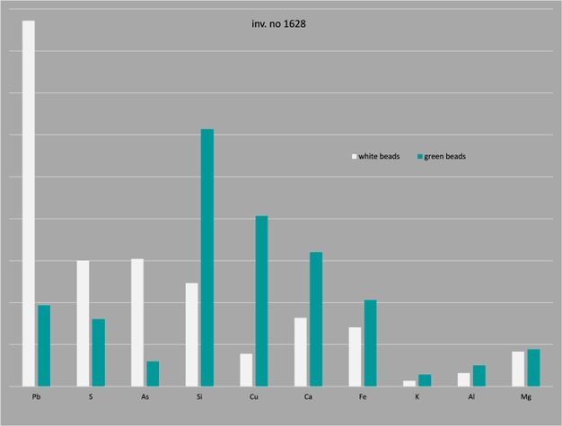
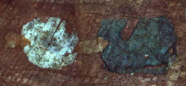
Although the citation of numerous XRF results exceeds the scope of this paper, a special mention must be given to the jewelry beads in the woman’s portrait, in relevance to their Egyptian blue content and how it relates to the VIL image. As previously noted, the white and the green-blue beads present a very similar, if not identical, degree of Egyptian blue luminescence in the IR spectrum. This finding raised the question of whether the proportions of Egyptian blue in the mixture of each colorant was of equal amounts. The elemental analysis performed with the XRF indicated that, when compared with the white beads, the green-blue beads have higher values of copper, silica, and calcium (the main compounds of Egyptian blue) as well as iron (fig. 5.20). The strong response of both the green-blue and the white beads with VIL suggests that the proportions of Egyptian blue present in the paint are not quantitative.
Microscopic Observation
Microscopy could be considered an imaging technique; however, as an examination tool, it complements the other procedures used, aiding significantly to our understanding of the portraits’ unique characteristics. Thus, microscopy should be mentioned separately.
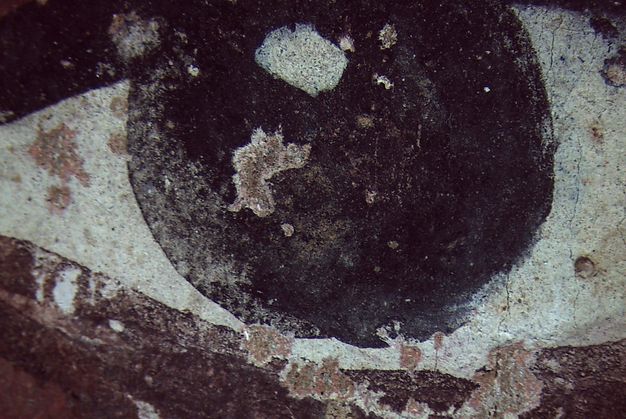
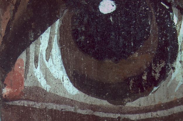
We used a Leica optical microscope equipped with a digital camera to examine as much of the surface as possible. Therefore, all the XRF target locations were compared with the microscopy performed at different magnifications. Both male portraits appear to have a very thin ground layer (or none at all) applied on the wooden support; however, they have distinct differences in the style and the layering of the features of the eyes (figs. 5.21 and 5.22). In the case of the young man, the round iris was put down first, followed by the white sclera around it. The eye of the older man was painted in the opposite manner: first the sclera, then the iris, followed by the brown semicircle to form the inner pupil and on top of them the white highlights of the sclera, and lastly the dark details.
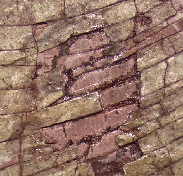
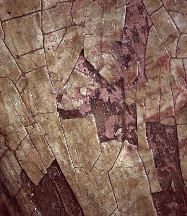
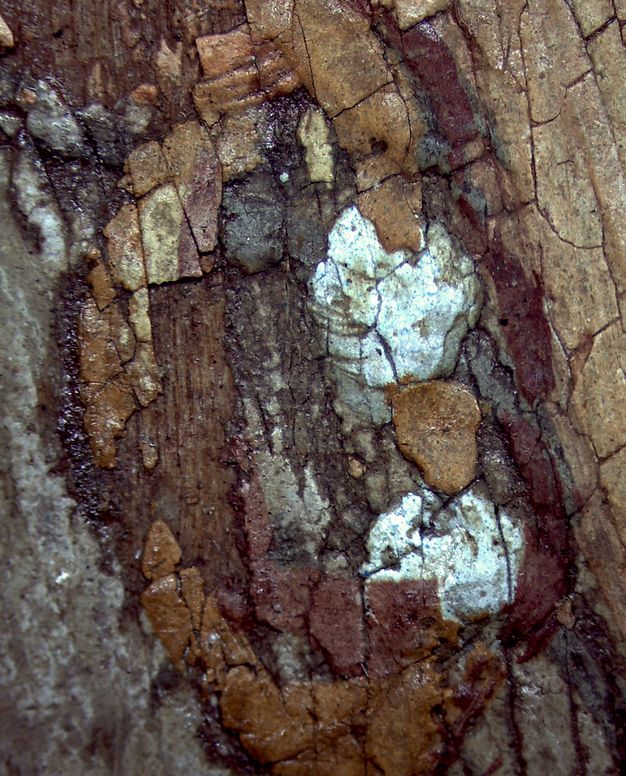
In the female portrait, it is obvious, even macroscopically, that the paint surface is significantly thicker than those of the other two portraits, and under the microscope it becomes even more apparent. Specifically, some areas of skin have lost their top layer, revealing a lower pink layer (figs. 5.23 and 5.24). That same thick layering of paint is also very evident in the jewelry, and the sequence of applying the details becomes more apparent—for example, in the “gold” hoop of the earring and the “gold” links of the necklace that overlap the beads (figs. 5.25 and 5.26).
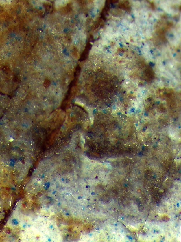
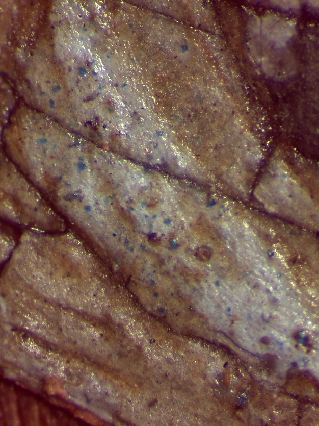
In terms of paint composition, various pigments were used in most instances. This fact is very clearly demonstrated in the microscopic images of a white bead on the woman’s necklace and of an outer corner highlight of her PR eye (figs. 5.27 and 5.28), where granules of Egyptian blue can be observed in the white paint.
Conclusions
This paper has described the scientific study of three funerary portraits in the Egyptian collection at the NAM, examined for the first time at the museum to overcome the challenge of “decoding” the portraits holistically. The methods used are part of the current infrastructure for non-invasive investigation of the Department of Conservation, Physical-Chemical Research, and Archaeometry at the NAM and were employed conjunctively.
In specific instances, XRF analysis provided the means to better understand the results of the imaging techniques for pigment identification in relation to the pigments’ unique characteristics. Specifically, we could better distinguish the pigments present in each portrait, as seen in figure 5.29, which enabled us to form robust deductions on the manufacture of the portraits and the intent of their creators. For example, the white beads of the woman’s jewelry most likely represent pearls and were painted using a mixture of mainly lead white, some orpiment, and a little Egyptian blue. The green-blue beads, however, most likely are meant to represent emeralds. According to the XRF results, the mixture used to achieve the precious stone’s color was Egyptian blue (also verified by VIL) with possibly a green pigment and the addition of some yellows and reds. The presence of arsenic (As) and mercury (Hg) (see fig. 5.19) strongly indicates the use of realgar or orpiment and cinnabar. The addition of Egyptian blue in the mixture also shows the artist’s attempt to imitate a specific type of highly valued emerald.8 The fact that the jewelry is carefully rendered suggests that the portrait was intended to portray a person of wealth or a high-ranking member of society.
| Colorant | No. 1627 (Young Man) |
No. 1628 (Woman) |
No. 1629 (Older Man) |
|---|---|---|---|
| Iron oxide (Umber or Ochre) | P | P | P |
| Orpiment or Realgar | P | P | P |
| Cinnabar | P | ||
| Gypsum | P | P | |
| Kaolin | P | ||
| Lead oxide | P | P | |
| Egyptian blue | P | ||
| Green earth | P | P | P |
| Malachite | ? | ||
| Red lake | P | P | |
| Indigo | ? | ? |
The same cannot be said for the production of the portrait of the young man. Here, the artist took a more minimal approach, and the color palette used seems to be rather poor. It looks as though the artist chose limited pigments and made no attempt to depict shadows and highlights.
Somewhere in the middle lies the production of the older man’s likeness. The sitter has some degree of detailing, such as the addition of shadows and highlights and more ornamentation (with the wreath). His color palette has been more carefully chosen, as is evident in the formation of the wreath and clavus. IRRFC images confirm that the artist used various pigments, such as those based on lead, arsenic, iron, and possibly green earth or indigo, for the wreath. As for the clavus, apart from madder lake, there is strong indication that realgar was also used.
Despite our initial understanding and subsequent investigation of these portraits—and the considerable new information that has been brought to light—some aspects still require investigation: the identification of the wood used for the panels and the analysis of the organic substances (colorants, varnishes, adhesives, binding media). The prospect of future work is exciting and challenging. We feel that the main benefits gained by our participation in the APPEAR project are the methods established at the NAM and the exchange of scientific investigations in the study of these precious artifacts.
Acknowledgments
We would like to thank the following individuals at the NAM: Dr. Konstantinos Nikolentzos, head of the Department of Prehistoric, Egyptian, Cypriot, and Near Eastern Antiquities Collection; Dr. Georgianna Moraitou, head of the Department of Conservation, Physical-Chemical Research, and Archaeometry; Panagiotis Lazaris, conservator in the Organic Materials, Paper, and Photo Negatives Conservation Laboratory; and Dr. Eleni Tourna, archaeologist in the Department of Prehistoric, Egyptian, Cypriot, and Near Eastern Antiquities Collection.
Notes
-
Doxiadis, Euphrosyne. 1995. The Mysterious Fayum Portraits: Faces from Ancient Egypt. Thames & Hudson.. ↩︎
-
Measday, Danielle, Charlotte Walker, and Briony Pemberton. 2017. “A Summary of Ultra-Violet Fluorescent Materials Relevant to Conservation.” AICCM National Newsletter, no. 137. Accessed April 26, 2021. https://aiccm.org.au/national-news/summary-ultra-violet-fluorescent-materials-relevant-conservation.. ↩︎
-
Digital processing using Photoshop CC 2015 by Adobe. ↩︎
-
Boust, Clotilde, and Anne Wohlgelmuth. 2017. “Database: Pigments under UV and IR Radiations.” Scientific Imaging for Cultural Heritage/Images scientifiques pour le patrimoine. Hypotheses, August 18. Accessed June 30, 2021. https://copa.hypotheses.org/552.. ↩︎
-
Boust, Clotilde, and Anne Wohlgelmuth. 2017. “Database: Pigments under UV and IR Radiations.” Scientific Imaging for Cultural Heritage/Images scientifiques pour le patrimoine. Hypotheses, August 18. Accessed June 30, 2021. https://copa.hypotheses.org/552.. ↩︎
-
In modern commercial terms, a rich bluish-green hue and a deep intense tone are considered characteristics of the more valuable emeralds. See Gemological Institute of America. n.d. “Buyer’s Guide.” Accessed September 9, 2022. https://www.gia.edu/emerald/buyers-guide. ↩︎