History of the Mummy Portrait
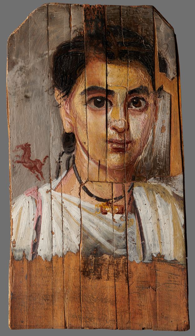
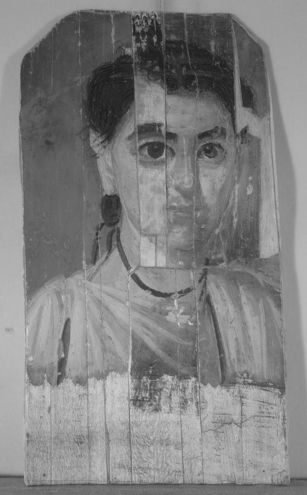
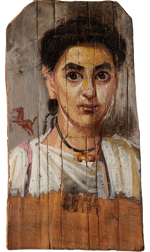
The only mummy portrait in the collection of the National Museum in Warsaw (NMW) (fig. 16.1) was purchased in Egypt, most likely in 1898, by eminent Kraków architect Józef Pakies (1858–1923), who acquired it from a museum in Giza.1 The work was kept at the Czartoryski Museum in Kraków. Georgius Werner included its first known photograph (fig. 16.2) in his article “De imaginibus Graeco-Aegyptis in colonia cui El-Fayum nomen est, repertis et Cracoviae asservatis observationes,” published in the Lviv periodical Eos in 1909. Werner recognized the representation as a girl and noted the significant damage to the board onto which the portrait was painted.2 In 1939, the work was acquired for the NMW from Dr. Władysław Kahl for 1,500 Polish zlotys, a modest sum both then and now (approximately $283 in 1939).
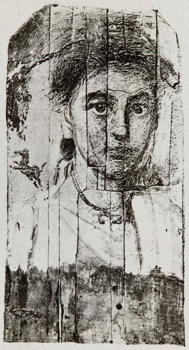
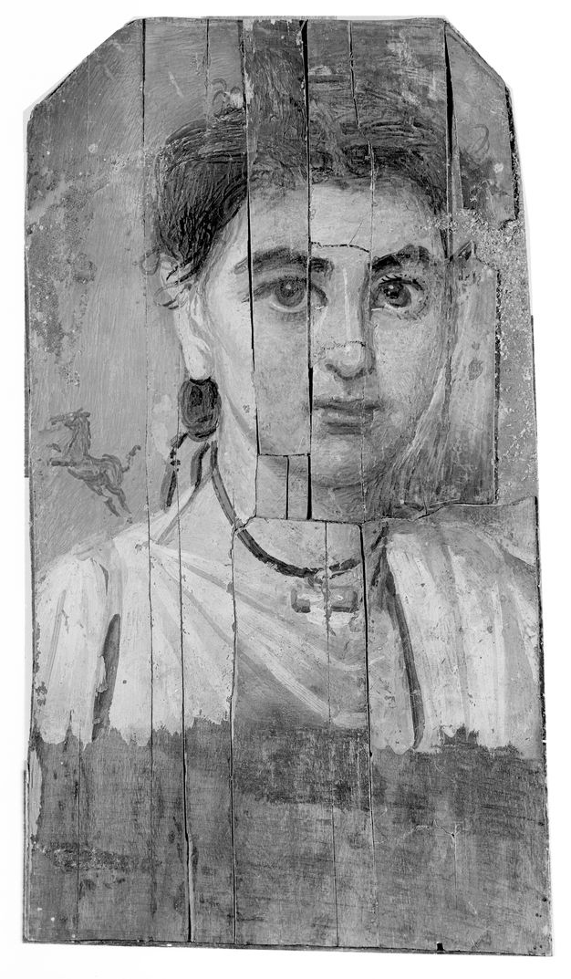
The portrait was entered into the museum’s inventory in May of the same year (the invoice, dated 22 May, also described the sitter as a girl). During World War II, the very existence of the object was seriously threatened. According to the account of prominent conservator Bohdan Marconi, it was “privately” stolen in late 1939 by Theo Diesel (SS-Untersturmführer, who led the confiscation of works of art at the museum in 1939–1940) and kept in Diesel’s room in the Bristol Hotel, but soon returned to the museum. Removed from Poland by the Nazis presumably in 1944, the work returned to the National Museum through restitution efforts. In October 1946, the portrait was discovered in one of the crates brought from Fischhorn Castle in Austria that contained items looted during the war.3 There is no documentation to show how storage conditions between 1939 and 1946 affected the portrait’s state of preservation. However, its condition in the 1946 post-war photo (fig. 16.3) is nearly identical to that in the 1909 photo. In his article published in 1966, archaeologist Janusz Ostrowski discussed the authenticity of several parts of the portrait.4 This was the first time that the work was described as depicting a boy.5
Description of the Portrait
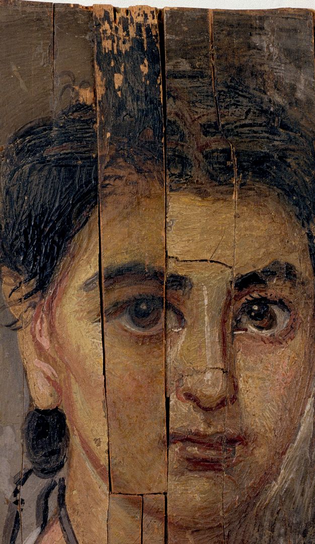
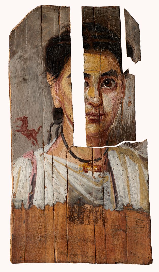
The portrait dates to the second half of the second century CE. It shows a youth’s face with regular features and large eyes looking straight ahead. His dark hair is pulled back and cinched behind the ear. The boy wears a white tunic with a vertical pink clavus over his right shoulder. On his neck is an amulet, a container for an apotropaic text, and above his right shoulder is a rearing horse painted in dark rose, a rare motif in funerary portraits whose meaning requires further research. The “lock of youth” and bulla, an amulet protecting the wearer from the evil eye, are the attributes of a boy of early age.6
The portrait appears to be painted in encaustic. The board is 37 x 18 cm and the upper corners are cut at an angle, which some scholars believe to be characteristic of the local tradition in er-Rubayat.7 The support is very thin, about 1–2 mm. The NMW laboratory identified the wood as linden after analyzing thin sections of wood through the optical microscope in transmitted light.8 The bottom of the board is left unpainted, with visible traces of a black layer. This thin black primer can also be detected in some places where the painting layer is slightly translucent.
For the most part, the paint was thickly and vividly applied in a well-practiced formula. The upper edges of the panel seem to have been cut after painting. Evidence for this is visible at the top of the panel, below where it was trimmed, where an incised line for marking (or guiding) the shaping of the panel is still evident in the paint. The background, which was likely painted first,9 outlines the figure, a practice also observed on other mummy portraits.10 Observations in raking light show a variety of textures and methods of paint application suggesting the use of brushes and other tools, possibly hard and pointed. Parallel brushstrokes are clearly visible on the painted surface of the gray background and, especially, on the boy’s white tunic. The face, neck, and hair texture reveals a modelling method that creates various patterns, likely the result of the use of metal tools in the paint’s application.11 Additionally, there are striking differences in highlights and shadows in the figure’s face and neck that give the work a beautiful, expressive appearance.
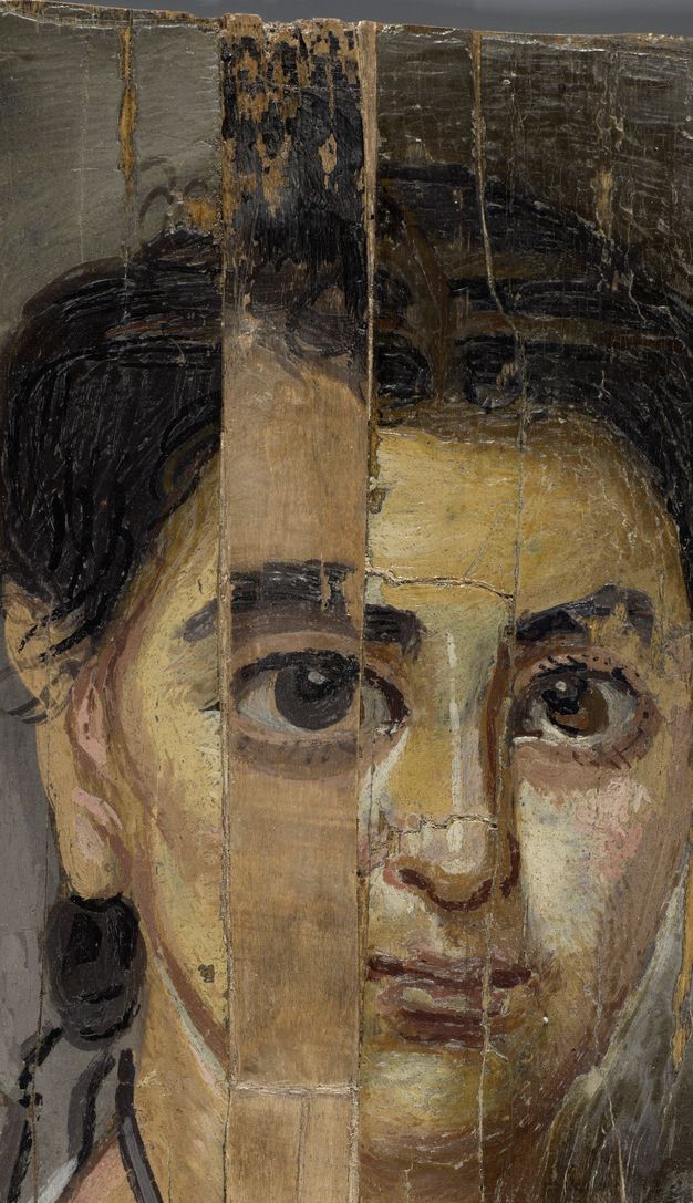
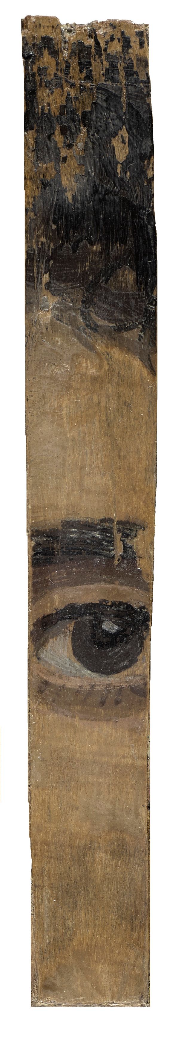
The panel is split both vertically and horizontally in several places and is slightly deformed and concave. Approximately nine separate pieces have been joined and glued to a cardboard backing. A vertical section that includes the boy’s right eye is a later addition—an element most likely originating from a similar ancient mummy portrait. The added fragment is raised above the surface because the left edge overlaps with the adjoining piece. The paint exhibits a craquelure pattern in irregular angular shapes in the white tunic and the gray background. In the darker areas, the cracks assume simple shapes and tend to be smaller. The blistering of the paint layer in the upper part of the pink clavus is consolidated with an adhesive. The wooden panel has deformed due to changes in relative humidity, causing movement of the split panel and the cardboard backing and resulting in a straight or bent concave shape (cradle) of both the entire portrait and the individual splits.
Non-Invasive Examination: Technical Imaging and Spectroscopy
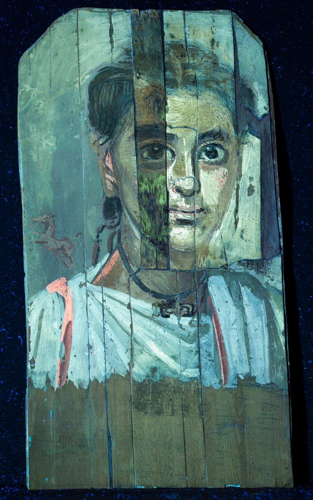
The portrait was examined using non-invasive technical imaging methods—ultraviolet-induced visible fluorescence (UVF), infrared reflectography (IRR), X-radiography, computed tomography (CT), X-ray fluorescence spectroscopy (XRF), and Fourier transform infrared spectroscopy (FTIR)—to gather more information about its overall condition. The images were made after dirt and overpaint removal. The research aimed to establish whether the paint layer is homogeneous and whether all of the sections mounted on cardboard come from the same object.
UV imaging (fig. 16.4)12 shows clear differences between the fluorescence of the paint layers on the fragment with the right eye and the rest of the composition. Other fluorescent areas include: a green color in areas of modern retouching; a slight difference in the luminosity on the left side of the face, which ends along the horizontal crack across the neck; a bright orange-to-pink area, characteristic of the type usually emitted by red lake pigments, in the area of the head and the tunic (on the clavus, evidence for the use of madder has been corroborated by FTIR analysis); and a light orange fluorescence above the right side of the hair and background, presumably a trace of shellac that was also confirmed by an analytical examination. Additionally, in the IRR image,13 the striking outline of the right eye, the eyebrow, and the hairline above the right eye is visibly different from the adjacent composition (fig. 16.5).
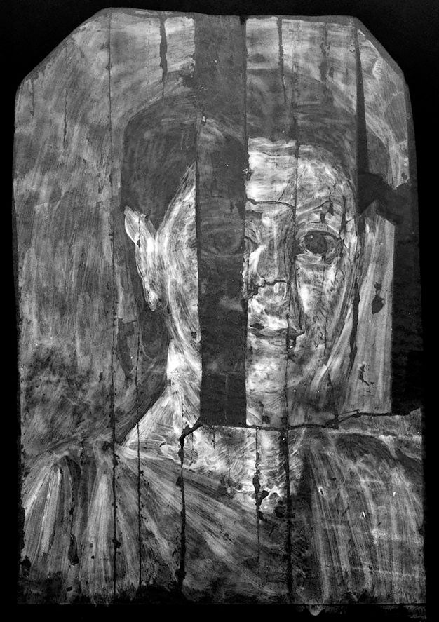
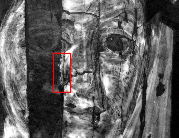
The portrait was further examined by X-radiography and computed tomography.14 X-ray images revealed information about the painting process and the uniformity of the paint layers. More importantly, X-radiography was able to detect the continuity of brushstrokes between separate split pieces (fig. 16.6). This was possible due to the strong X-ray absorption of lead pigments used in the paint layers, identified during analytical testing. Technological differences between the fragment with the right eye and the rest of the portrait were also confirmed. The hidden fragment of the panel under the board with the right eye was visualized along with the irregular shape (line) of the damaged edge of the panel at this location (fig. 16.7). Computed tomography enabled us to penetrate the work, making it possible to recognize the state of preservation, structure, and materials, and retrace the artist’s creative process.15 The interpretation of the obtained multi-layered cross section is still pending analysis.
Microscopic observation brought several important findings to light that will require further research. The surface of the encaustic paint of the portrait was especially varied in terms of composition and texture. We documented some deposits inside the paint, likely connected with the process of painting with encaustic. A piece of reed or wood was noticed, alongside numerous short black hairs and one fragment of a red thread. Cavities from air bubbles were also observed on the surface.
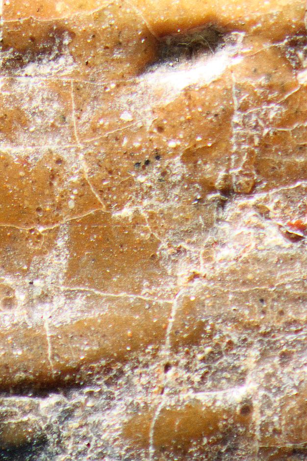
Another finding was the observation of the white particles firmly attached to the paint surface. These individual white deposits, resembling scattered crumbs, were visible on the surface of the gray background (fig. 16.8). Different minor white spots in the form of clusters were noticed on the right side of the painting (fig. 16.9) (see following section).
Pigment Investigation and Elemental Analysis Using Portable X-Ray Fluorescence Spectroscopy (p-XRF)
All measurements were performed using an XRF Tracer III-SD spectrometer (Bruker) equipped with a silicon drift detector (SDD detector).16 The system enables elemental analysis in the Mg–Pu range. A primary X-ray radiation lamp with an Rh anode with variable 2–25 μA current and maximum voltage up to 45 kV was used. All XRF spectra were acquired from areas of ca. 0.5cm2, and no subtle variabilities between the analyzed areas could be detected. The following instrumental settings and data acquisition parameters were used: voltage 45 kV; current 23.1 μA; spectra acquisition time 60 s; pressure < 60 Tr.
XRF spectra were registered using S1PXRF software and then compiled in Excel. The presence of Pb was detected in all samples. The presence of barium was also detected in all analyzed areas by recognition of BaK⍺1 at 32.19 keV, BaK⍺2 at 31.82 keV and BaKβ1 at 36.38 keV. The information about Ba versus Ti was selected after careful examination of the presence of their signals at the characteristic energies (TiK⍺1/K⍺2 at 4.50/4.51 keV; TiKb1 at 4.93 keV; BaL⍺1/ L⍺2 at 4.47/4.45 keV; and BaLβ1 at 4.83 keV), bearing in mind that they are difficult to distinguish while using p-XRF. The highest Zn signals accompanied Ba in the same areas. The interpretation of these results is not obvious, but barite (BaSO4) is a relatively common mineral. It is typically found as an opaque white heavy mineral in sedimentary rocks such as limestone. It can be also associated with sulfide secondary minerals, like galena (PbS) or sphalerite (ZnS). It is possible that the mineral barite might be used as a filler in the painting.
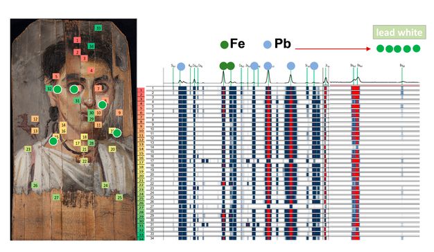
The XRF results allow us to make some conclusions about the presence of lead white (basic lead carbonate) and iron oxides in brown, yellow, and red (lips) areas. The red horse was probably painted with organic pigments, which could not be identified by means of XRF (fig. 16.10).
Organic Analysis Using Fourier Transform Infrared Spectroscopy (FTIR)
The spectra obtained from the samples were recorded by attenuated total reflection Fourier transform infrared (ATR-FTIR) spectrometer (Alpha, Bruker Optics, Germany USA-MA) equipped with a diamond crystal. The spectra were collected in the 4000–400 cm-1 range with a resolution of 4 cm-1. The characteristic bandwidths were interpreted and compared with literature data17 and reference spectra collected in the laboratory.
One microsample was taken for analysis by FTIR-ATR spectrometry. The material was collected from an area on the pink clavus (near the lower edge of the painting from an area of loss) where we suspected the presence of an organic dye. Peaks characteristic of wax (2918, 2846, 1736, 1457, 1169, and 721 cm-1) were visible in the sample’s spectrum. Other peaks at 1091, 721, 674, and below 600 cm-1 were also identified. Their presence resembles spectra of a plant-based red containing anthraquinone, a commonly used pigment in antiquity and a component of madder lake.18
A second microsample from the black ground layer was taken to identify organic binder. The spectra with characteristic peaks at 2918, 2851, 1631, 1421, and 1379 cm-1 correlate a reference spectrum for bitumen, also commonly used in antiquity in the production of mummified human remains.19
Other Investigations Using FTIR-ATR
We used an FTIR-ATR spectrometer to examine a sample of the single white deposit from the gray background layer, comparing the results with the different-looking clusters of white spots within the brown background layer. Wax was identified in the sample taken from the gray area at the left edge near the boy’s shoulder. The peaks at 2922, 2851, 1736, 1461, 1167, and 718 cm-1 correspond to the reference spectra for beeswax. The presence of lead white, evidenced by peaks at 1403 and 681 cm-1, was also identified. Consequently, the results obtained for this sample aligned with the measurements taken with the portable spectrometer. An unusual signal appeared at 1515 cm-1, which did not correspond to any pure standard reference samples typically used in ancient Egypt. Signals recorded for the brown sample from the area of the upper edge (2920, 2849, 1738, 1463, 1421, 1171, 1026, and 723 cm-1) correlate with the reference spectrum for resin, most likely shellac from previous conservation treatments, confirming observations made with UV radiation. There is also a signal at 1519 cm-1, similar to the gray area sampled from the left panel edge in the boy’s shoulder.
Peaks between 1550 and 1510 cm-1 come from amide II band stretching, mainly showing N-H motion. These signals are attributed to protein deformation in the literature concerning protein interactions with pigments20 and the investigation of funerary masks.21 In the case of the current portrait, they may also indicate either the use of egg or reactions caused by a series of factors such as the presence of fungi or an interaction with substances used in the mummification process. Experiments concerning reactions with fungi have already been explored but need to be continued, considering the artificial aging process in such a complex environment.
FTIR Binding-Media Analysis
The binding media used to paint the NMW’s mummy portrait were identified using FTIR. This method allows the determination of the material groups used and the identification of possible interventions to the object. The analysis was performed using an ALPHA (Bruker) portable spectrometer equipped with a module for non-invasive analysis. The module, with 4 mm diameter openings, was placed against representative points on the surface of the painting. The selected spots were analyzed with the use of a transmitted infrared beam. Consequently, we were able to obtain molecular information about a wide range of painting materials. The characteristic bandwidths produced by individual bonds and functional groups were also identified.
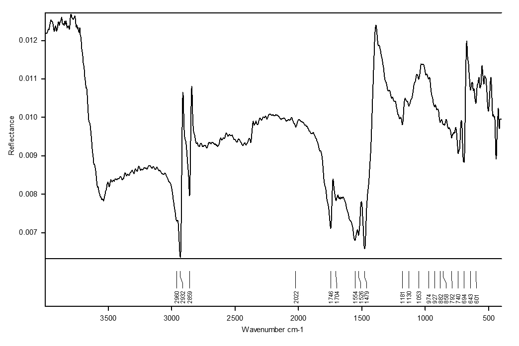
The exemplary FTIR reflectance spectra from the skin area at the center of the proper right cheek are shown in figure 16.11. The presence of wax (2932 cm-1, 1746 cm-1, and 1704 cm-1), oil (2960 cm-1), protein (1554 cm-1), and lead white (1479 cm-1, 694 cm-1) was identified. It was possible to identify the presence of wax and protein in the nine analyzed points. Oil was identified in all areas except the temple near the proper right ear. The presence of lead white, whose high-intensity signals often make it difficult to distinguish bandwidths characteristic of other substances, interfered with the analysis.
Conservation
The portrait, cracked and broken into individual narrow sections, was most likely mounted onto cardboard when it was still in Egypt. It is also likely that this was when an antiquities dealer replaced a destroyed fragment of the right eye with a section originating from another mummy portrait. There is no evidence of conservation treatment between the time that the portrait was acquired for the NMW and 1998, when an analytical report was published describing the reduction of a white bloom that appeared on the painting’s surface. The decision was made to stabilize the portrait in 2004, when the paint layer began to flake and crack and the portrait’s cardboard backing continued to detach from the panel. During the conservation treatment, dirt and overpaint were removed from the surface of the painting, the flaking paint layer was consolidated, and the cardboard was stabilized. Even before conservation treatment, in 2005,22 there were visible differences between the portrait’s fragments (fig. 16.12). After the surface was cleaned, the section containing the proper right eye appeared more distracting. It was determined that this area had been overpainted to hide various anomalies: the eye painted on the board that was originally larger, the brow was higher, and the hairline inconsistent with the rest of the portrait (fig. 16.13). The examination also proved the use of different techniques for the added section.
After the removal of dirt and overpainting, the portrait’s new appearance was poorly received by viewers. However, the decision was made against changing the addition of later sections or removing the cardboard backing, considering that such manipulations could damage the paint layer and wood.23 Following the curatorial request, the inserted section was retouched with (reversible) watercolor paint in the form of minute tratteggio (hatching) lines to minimize the visual difference between the added piece and the rest of the composition.
Following our investigation, a digital rendering of the presumed original appearance of the portrait was made. The images of matching pieces (fig. 16.14) as well as the separate section with the eye (fig. 16.15) were prepared based on the assumption that the latter probably originated from another mummy portrait, and a hypothetical reconstruction of the portrait (fig. 16.16) was made. According to the observations, neither the shape nor the size of the transplanted section fit in with the rest of the work and this is why its left edge is slightly raised on the neighboring board. We noticed that the edge where the portrait cracked horizontally was inconsistent in color with the neighboring paint layers. This inconsistency is visible in the imaging examination (see figs. 16.6 and 16.7). Additionally, the vertical cracks do not run in one line, suggesting that the left side of the portrait should be shifted toward the center, and a small fragment on the left edge was not positioned correctly. In our digital reconstruction, we corrected the position of the sections and made a color reconstruction of the missing part. Fig. 16.16 shows a completed face of the boy and, additionally, a separate fragment of another portrait (see fig. 16.15).
Observation Summary
The wood panel was identified as linden over the course of our research. The vertical section with the boy’s proper right eye (see fig. 16.15) was recognized as an element most likely originating from another ancient mummy portrait. UVF, X-radiography, and IRR show clear differences between the response of the paint layers on this added fragment and the rest of the object. X-radiography provided information about the painting process and the uniformity of the paint layers, allowing us to detect the continuity of brushstrokes between the other separated sections. Wax, oil, lead white, earth compounds, madder, and bitumen were detected using a variety of analytical methods. Based on the XRF results the presence of lead white (basic lead carbonate) and iron oxides in the brown, yellow, and red (lips) areas is suggested.
Conclusions
The materials identified during the examination of the object are consistent with mummy portrait production in second-century Egypt. The portrait from the NMW may come from one of the known Egyptian sites24 (based on the shape of the original panel, possibly er-Rubayat) and made its way to Giza before being purchased by the Polish architect Józef Pakies. The object’s condition, combined with our investigations and analyses, prove that the portrait underwent conservation treatment before 1909, when it was photographed and analyzed by Georgius Werner. Fragments of the cracked portrait of the boy were mounted on cardboard and the damaged part of the face was reconstituted from an ancient panel removed from a different mummy portrait. It was then overpainted to unify the added board with the rest of the composition. The artifact could have been part of a group of works discovered in Egypt and restored for the antiquities market at the end of the nineteenth century by creating a pastiche of better-preserved sections of similar portraits.25 In recent years, following the removal of the modern overpainting, tratteggio was used to unify and improve the appearance for display in the Ancient Art Gallery in the National Museum in Warsaw.
Notes
-
Werner, Georgius. 1909. “De imaginibus Graeco-Aegyptis in colonia cui El-Fayum nomen est, repertis et Cracoviae asservatis observations.” Eos 15: 130–37.. Fig. 16.1: Inv. no. 23676 MNW (former inv. no. 127191 MNW). ↩︎
-
Werner, Georgius. 1909. “De imaginibus Graeco-Aegyptis in colonia cui El-Fayum nomen est, repertis et Cracoviae asservatis observations.” Eos 15: 130–37.. ↩︎
-
Marconi, Bohdan. 1967. “Wspomnienia z lat 1939–1945.” Rocznik Muzeum Narodowego w Warszawie 11: 269–77.. Maria Ludwika Bernhard, “Sprawozdanie do Kroniki za mc. październik 1946 r. Zbiory Sztuki Starożytnej” (The Report for the Chronicle to October 1946). The Archive of the National Museum in Warsaw, no. PL/304/1/0/859, 132. We are grateful to Kacper Laube (Department of Ancient Art, National Muzeum Warsaw) for turning our attention to this archival document; see also Bernhard, Maria Ludwika. 1947. “Zbiory Sztuki Starożytnej w Muzeum Narodowym w Warszawie w latach 1939–1946.” Archeologia 1: 302–9., and Michałowski, Kazimierz. 1957. “Galeria Sztuki Starożytnej w Muzeum Narodowym w Warszawie.” Rocznik Muzeum Narodowego w Warszawie 2: 101–38.. ↩︎
-
Ostrowski, Janusz. 1966. “Portret fajumski ze zbiorów Muzeum Narodowego w Warszawie.” Rocznik Muzeum Narodowego w Warszawie 10: 13–22.. ↩︎
-
Ostrowski, Janusz. 1966. “Portret fajumski ze zbiorów Muzeum Narodowego w Warszawie.” Rocznik Muzeum Narodowego w Warszawie 10: 13–22.. ↩︎
-
Laes, Christian, and Johan Strubbe. 2014. Youth in the Roman Empire: The Young and the Restless Years? Cambridge University Press.; Montserrat, Dominic. 1993. “The Representation of Young Males in ‘Fayum Portraits.’” The Journal of Egyptian Archaeology 79: 215–25.; Wiedemann, Thomas. 2014. Adults and Children in the Roman Empire. Routledge.. ↩︎
-
Doxiadis, Euphrosyne. 1995. The Mysterious Fayum Portraits: Faces from Ancient Egypt. Thames & Hudson.; Freccero, Agneta. 2000. Fayum Portraits: Documentation and Scientific Analysis of Mummy Portraits Belonging to Nationalmuseum in Stockholm. Acta Universitatis Gothoborgensis.. ↩︎
-
Tilia sp. The wood was identified by Iwona Pannenko in 2006. ↩︎
-
van Daal, Jan. 2019. “Ancient Incarnations: Depicting Human Flesh in Mummy Portraits from Roman Egypt.” Master of Science thesis, University of Amsterdam.. ↩︎
-
Thompson, David L. 1982. Mummy Portraits in the J. Paul Getty Museum. J. Paul Getty Museum.; Salvant, Johanna, Jane Williams, Monica Ganio, Francesca Casidio, Céline Daher, Ken Sutherland, Letizia Monico, Frederik Vanmeert, Steven de Meyer, Koen Janssens, Caroline R. Cartwright, and Marc Walton. 2018. “A Roman Egyptian Painting Workshop: Technical Investigation of the Portraits from Tebtunis, Egypt.” Archaeometry 60 (4): 815–33. https://doi.org/10.1111/arcm.12351.. ↩︎
-
Bostock, J., and H. Riley, trans. 1855. The Natural History (Naturalis Historia). By Pliny the Elder. Taylor and Francis; Henry G. Bohn., XXXV, 41. ↩︎
-
Canon 5D Mark II camera, Canon Macro fixed focal 100 mm lens, UV wavelength 300–400 nm. Image by Anna Lewandowska. ↩︎
-
Canon 6D camera, modified to IR wavelength range of above 1000 nm, Canon EF 50 mm f/1.8 lens, 830 nm and 1000 nm filters. Image by Anna Lewandowska. ↩︎
-
The painting was imaged using a Philips Digital Diagnost X-ray system and Neusoft NeuViz 64 En 64-slices computed tomography scanner, thanks to a collaboration between the NMW and the European Health Centre Otwock (EHC). The imaging was performed by Łukasz Kownacki, MD, PhD, in the Department of Diagnostic Imaging, EHC, in 2017. ↩︎
-
As part of a long-term project between the NMW and the EHC directed by Łukasz Kownacki and Dorota Ignatowicz-Woźniakowska (senior conservator for specialized diagnostic imaging at the NMW) in cooperation with scientists working in this area of research. ↩︎
-
We would like to thank Olga Syta, PhD, for her contribution to the XRF investigation. ↩︎
-
Zaffino, Chiara, Vittoria Guglielmi, Silvio Faraone, Alessandro Vinaccia, and Silvia Bruni. 2015. “Exploiting External Reflection FTIR Spectroscopy for the In-Situ Identification of Pigments and Binders in Illuminated Manuscripts. Brochantite and Posnjakite as a Case Study.” Spectrochimica Acta: Part A, Molecular and Biomolecular Spectroscopy 136 (B): 1076–85.; Rosi, Francesca, Laura Cartechini, Diego Sali, and Costanza Miliani. 2019. “Recent Trends in the Application of Fourier Transform Infrared (FT-IR) Spectroscopy in Heritage Science: From Micro- to Non-Invasive FT-IR.” Physical Sciences Reviews 4: n.p.; Hofko, Bernhard, Mohammad Zia Alavi, Hinrich Grothe, David Jones, and John Harvey. 2017. “Repeatability and Sensitivity of FTIR ATR Spectral Analysis Methods for Bituminous Binders.” Materials and Structures 50 (3): 1–15.. ↩︎
-
Scott, David A. 2016. “A Review of Ancient Egyptian Pigments and Cosmetics.” Studies in Conservation 61 (4): 185–202. https://doi.org/10.1179/2047058414Y.0000000162.; Nicholson, Paul T., and Ian Shaw. 2000. Ancient Egyptian Materials and Technology. Cambridge University Press.. ↩︎
-
Scott, David A. 2016. “A Review of Ancient Egyptian Pigments and Cosmetics.” Studies in Conservation 61 (4): 185–202. https://doi.org/10.1179/2047058414Y.0000000162.; Nicholson, Paul T., and Ian Shaw. 2000. Ancient Egyptian Materials and Technology. Cambridge University Press.. ↩︎
-
Duce, Celia, Lisa Ghezzi, Massimo Onor, Ilaria Bonaduce, Maria Perla Colombini, Maria Rosaria Tine’, and Emilia Bramanti. 2012. “Physico-Chemical Characterisation of Protein-Pigment Interactions in Tempera Paint Reconstructions: Casein/Cinnabar and Albumin/Cinnabar.” Analytical and Bioanalytical Chemistry 402 (6): 2183–93.; Socrates, George. 2001. Infrared and Raman Characteristic Group Frequencies. Tables and Charts. 3rd ed. John Wiley & Sons.. ↩︎
-
Schiavon, Nick, Carlo Emmanuele Bottaini, Sara Valadas, Cristina Barrocas Dias, Ana Manhita, and Antonio Candeias. 2021. “A Multi-Analytical Study of Egyptian Funerary Artifacts from Three Portuguese Museum Collections.” Heritage 4 (4): 2973–95.. ↩︎
-
The cleaning and adhesive lining were done by Agnieszka Kijowska from the NMW Ancient Art Conservation Workshop. ↩︎
-
In 2005, we discussed the risks of removing the secondary support by email correspondence with conservators specializing in the subject of mummy portraits: Marie Svoboda (J. Paul Getty Museum, Los Angeles), Jane Williams (Phoebe A. Hearst Museum of Anthropology/Fine Arts Museums of San Francisco), Britta Nilsson (Nationalmuseum, Stockholm), and Nicola Newman (British Museum). ↩︎
-
Aubert, Marie-France. 2000. “Portraits from Antinoopolis and Other Sites.” In Walker 2000A.. ↩︎
-
Thompson, David L. 1981. “A Lost Patchwork ‘Fayum Portrait.’” American Journal of Archaeology 85 (4): 491–92.; Thompson, David L. 1973. “A Patchwork Fayum in Toledo.” American Journal of Archaeology 77 (4): 438–39., tab. 88. ↩︎