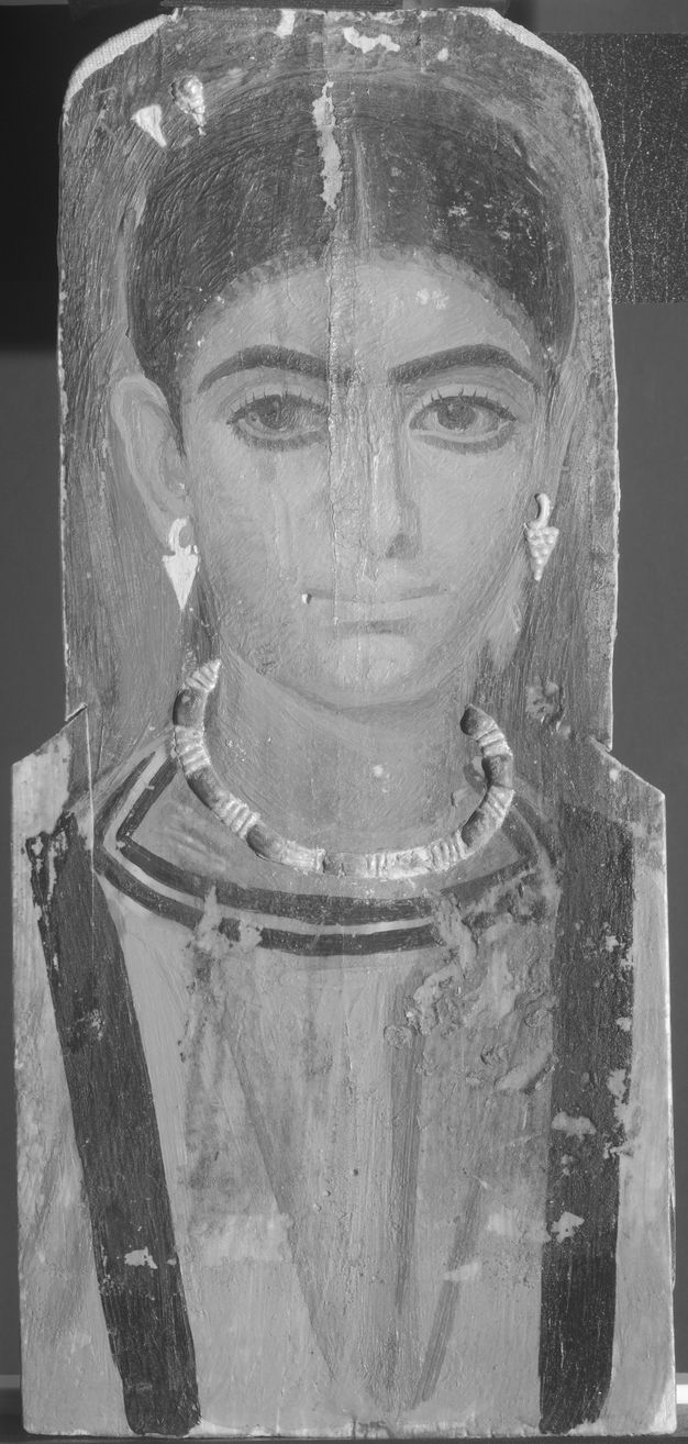
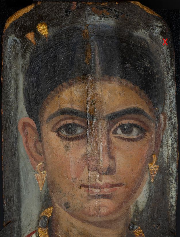
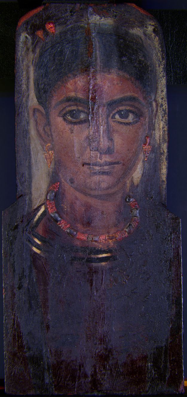
The collection of the Nelson-Atkins Museum of Art contains a Roman Egyptian funerary portrait from Antinoöpolis stylistically dated to the second century CE. Acquired in 1937, this encaustic painting on wood panel is representative of a corpus of funerary portraits painted during the Roman rule of Egypt from the first through third centuries CE. The portrait depicts a young woman with an austere expression, dressed in a deep red tunic with a black-and-white striped collar (or undertunic), and black clavi at the shoulders (fig. 14.1). The sitter is set against a dark gray background that transitions to a light gray highlight directly behind her head. The woman’s large, almond-shaped eyes are accentuated by individually painted lashes, and thick, arched eyebrows. Deep shadows and bright highlights on her olive complexion emphasize her strong features. Her jewelry, rendered in relief using gilt stucco, includes pendant earrings in the form of grape clusters, a gold necklace inset with dark blue stones modeled in painted stucco, and two hairpins that secure hair piled tightly atop her head in a thick plait. Multiple application techniques can be observed, including the marks of a sharp, pointed instrument used to render complex shading and delicate details of the face, neck, and head, while wide, vertical brushstrokes were used in the garment and background.
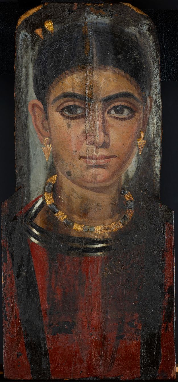
One gilt stucco hairpin and the right earring are lost, revealing the wood support and stucco remnants. The necklace’s painted blue stones are darkened by accretions and dirt that visually obscure the original color. The painted surface of the stone located to the proper left of the central stone is lost, leaving behind broken stucco remnants. The central blue stone in the necklace is cracked down its center, correlating to the vertical split through the center of the wood panel that was mended prior to painting and repaired by modern intervention. A clear coating applied along the central split below the large loss in the woman’s hair has darkened the paint surface from the forehead to the right nostril. The paint surface near the bottom of the panel is slightly sticky, with dust fibers stuck to the surface.
There are cracks in the encaustic surface throughout the portrait. The most significant paint losses are located at the top of the panel around the central crack and throughout the garment. Blanching of the paint surface has occurred in the dark background, namely along the proper left side, in smaller portions of the hair, and along the upper proper right side. Black surface deposits and staining visibly obscure a large portion of the sitter’s garment and extend into the neck. The paint surface hidden below these accretions on the proper left side is damaged with some paint loss. The paint is smeared around the collar and tunic, suggesting the wax was in a softened state when this damage occurred. The black deposits terminate around the outer edges of the clavi, several inches above the bottom of the panel, suggesting the outer edges of the panel were somehow protected from these deposits. Although there is no visual evidence of textile on the front, the well-preserved edges may indicate where the linen wrappings once secured the panel in place over the head of the mummified remains.
All that is known of the painting’s provenience is based on art historical critique from visual examination. The shape of the Nelson-Atkins panel, which lacks all outer remnants of the mummy wrappings, exhibits stepped shoulders that have been recognized as a unique characteristic of works from Antinoöpolis.1 Portraits from Antinoöpolis are cited for the depiction of solemn subjects wearing austere fashion, tightly confined hair styles, and include women often swathed in drapery.2 Scholars have narrowly attributed the portrait to the early Antonine period (131–160 CE) based on her hairstyle, which is parted down the center and piled atop her head in a thick bun with a line of fine curls at the forehead.3
This paper focuses on a recent technical study that attempted to broaden our understanding of this encaustic work by clarifying the artist’s working techniques and material choices not accessible through visual assessment alone. As more portraits are added to the Getty’s APPEAR (Ancient Panel Paintings: Examination, Analysis, and Research) project database, comparison of the Nelson-Atkins’s single example of this type of portrait with those from other collections may contribute to a better understanding of the group.
Methods
Several non-sampling examination techniques were employed to investigate the materials, composition, and working techniques of the portrait, followed by microanalyses of a small number of samples. The structure, condition, and extent of restoration were investigated without sampling by X-radiography, multiband imaging (MBI), and surface microscopy. MBI employs technology to observe an object in wavelength ranges that extend beyond the capabilities of the human eye, from the ultraviolet (UV) to the near infrared (NIR).4 Images were captured using a UV-VIS-IR-modified Nikon D7000 DSLR camera with a 60mm lens and a set of UV-VIS-IR bandpass filters. While the imaging set was comprehensive, only those images with diagnostic relevance will be discussed.
Elemental mapping by X-ray fluorescence spectrometry (XRF) was conducted on the painting using a small, portable instrument built in-house following a design provided by collaborators from the Laboratoire d’archéologie moléculaire et structurale–Centre national de la recherche scientifique (LAMS-CNRS), Paris, that has been previously published.5 Our instrument differs from the published design only in its use of a machined aluminum coupling component that offers improved heat dissipation for the detector’s electronics and two-axis motion in only X and Y directions. The essential features and operating conditions of the instrument include a Moxtek end-window X-ray tube with Pd anode, operated at 40kV and 50µA, and an Amptek X123 SDD detector with 25mm2 area. The tube output is collimated by a 0.7mm diameter Pd tube yielding an effective spot size of 1mm at the standoff distance of 15mm. Maps were acquired in two rectangular runs overlapping in the region where the panel width increases, one for the face and the other for the torso. The face was acquired over a 170 x 240mm area at 1mm spacings using dynamic acquisition (on the fly) with an acquisition interval of 0.5 seconds per spectrum. The torso acquisition was done over 205 x 210mm with a longer dwell time of 0.75 seconds. Acquisition control was programmed in Python and spectra were fitted using PyMca.6 Elemental maps output with 32-bit grayscales were rescaled to 8-bits for reproduction as standard graphics.
Microsamples from the red tunic and hazy background deposits were analyzed by polarized light microscopy (PLM) and scanning electron microscopy coupled with energy-dispersive X-ray spectrometry (SEM-EDX).
Results and Discussion
Egyptian Blue
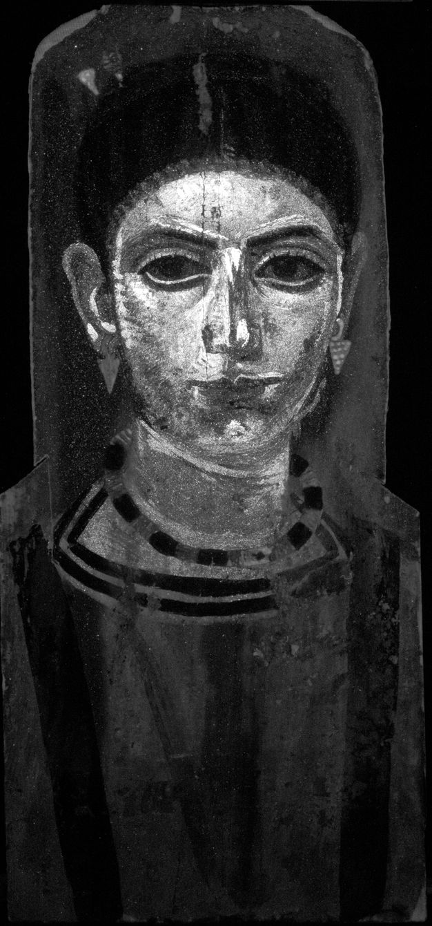
Egyptian blue, the synthetic blue pigment composed of copper calcium silicate, exhibits unique and intense photoluminescence that provides a complementary method to reflectance spectroscopy. The strong photo-induced near-infrared (NIR) luminescence emission at 910nm has been used as a characteristic marker to identify and map Egyptian blue in ancient paintings using visible-induced infrared luminescence (VIL) photography.7 Egyptian blue was initially discovered in the painting with VIL, showing sharply defined strokes contouring the figure’s eyes, brow line, head, and neck (fig. 14.2). XRF elemental mapping showed copper concentrations in the same areas of the face and neck, confirming the use of Egyptian blue in the painting (fig. 14.3A) while also disclosing copper in another form in the dark blue stones of the necklace. The stones are rendered in stucco relief and painted with a second copper-containing pigment that has not been identified in this study. Although the painting does not appear to visually employ Egyptian blue in the facial features, VIL and XRF uncovered the artist’s use of the blue pigment in two distinct roles: (1) as a means of sketching the underdrawing and (2) in the modulations of flesh tones in the upper-layer stratigraphy.
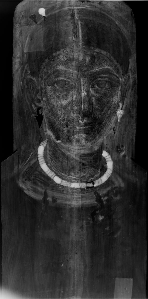
The copper response in the facial areas does not consistently correspond to either highlights or shadows but also seems to have been a means of laying out the proportions of the face as an underdrawing. A noticeable feature of the Egyptian blue distribution is the peak in each brow line that, together, form an “M.” An X-radiograph of the portrait shows the density associated with the heavier Egyptian blue applications that appear to be sketch lines above the eyes and along the nose (fig. 14.4). While lead white is also present in the face and contributes to the total radiographic density, the Egyptian blue application correlates to locations of increased density in the radiograph. When comparing the VIL image and copper map to the visible light image, the lines of Egyptian blue are in close proximity to the highlight above the eyebrows containing lead. Several blue particles are visible on the surface of the highlights when viewed under magnification, indicating the presence of some Egyptian blue in the paint mixture. A direct overlay of the elemental maps for copper and lead shows, however, some offset of the areas of maximum intensity for the two elements in the eyebrow and nose constructions. The lines for Egyptian blue fall slightly below and to the left of surface highlights that correspond to the lead map above the eyebrows and along the nose, respectively.
Underdrawings detected in a small number of portraits to date have more often relied on carbon black or madder than on Egyptian blue.8 The prominent role of Egyptian blue in this painting and its use in sketchwork are not apparent in normal viewing. In the absence of historical accounts of artists’ techniques from the period, a possible explanation could be that the pigment’s coarse texture allowed the sketch to remain perceptible beneath successive paint applications as an increased roughness until the work was nearly complete. A portrait of a woman excavated from the site of Tebtunis in the collection of the Phoebe A. Hearst Museum of Anthropology (PAHMA #6-21375) shows another example of Egyptian blue identified in the face that appears to be employed as an underdrawing, also discovered through VIL and complementary XRF methods.9 Much like that of the Nelson-Atkins’s sitter, VIL reveals Egyptian blue in outlines along her face and on the “M” above the sitter’s eyebrows.
The more common role of Egyptian blue—mixed with other pigments to enhance their colors10—is also seen in the Nelson-Atkins’s portrait. Egyptian blue was used to modulate flesh tones in both highlights and shading around the eyes, ears, nose, neck, and forehead. While a blue hue is imperceptible in these areas, examination of the paint surface under a stereomicroscope revealed particles of Egyptian blue in corresponding areas of highlights and shading as suggested in the VIL image and copper distribution map (fig. 14.5). Along with unprovenienced examples, portraits from Hawara and Saqqara have been found to utilize Egyptian blue in this manner.11 A VIL study of Egyptian blue in Roman Egyptian funerary portraits from the Cantor Arts Center at Stanford University12 confirmed the pigment was used in idiosyncratic ways in the flesh tones, garments, and eyes, but its use for underdrawing is noticeably absent from those findings.
Umber
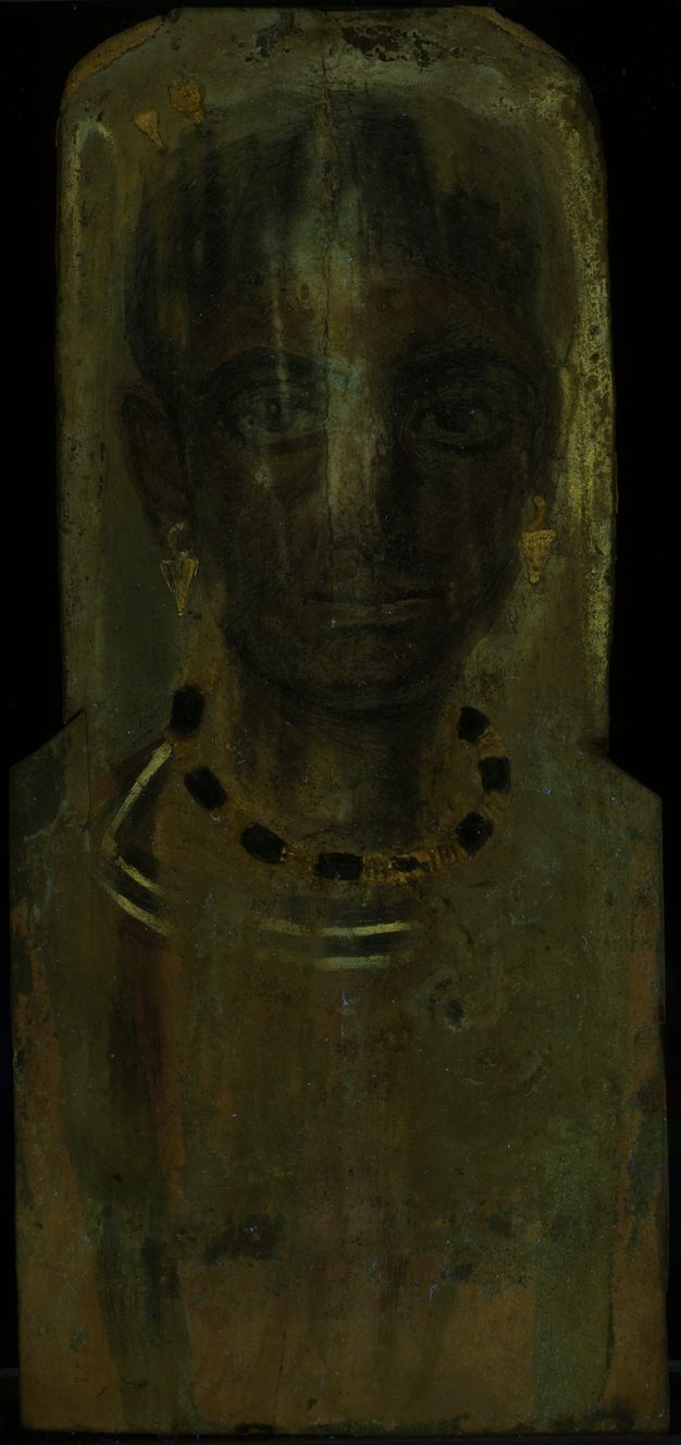
Elevated levels of manganese in dark areas of the painting were identified with elemental mapping. Manganese is elevated in shadows of the garment folds and shading of the facial features, both presumably using some form of umber (fig. 14.3B). Umber is known to be used in varying proportions with iron-earth pigments, carbon black, and lead white to achieve variations in brown tones in mummy portraits.13 A reflected infrared (IRR) image serves to differentiate nuances in dark brown and black passages when compared to the distribution of manganese derived from XRF mapping by revealing the distribution of carbon blacks not detectable with XRF (fig. 14.6). The IRR image demonstrates that carbon black was utilized in areas such as the hair, eyes, eyebrows, clavi, striped collar, and dark shading in the tunic.
It is clear from this comparison that manganese and carbon black play distinct roles in the depiction of dark details in the portrait. The hair is painted overall with carbon black, iron, and a small amount of manganese. The central “V” folds and the drapery on the proper right sleeve rely heavily on manganese and very little carbon black, while the clavi on the garment are treated differently on the right and left. The proper right clavus contains more manganese than the proper left, even though the two seem to contain equivalent applications of carbon black, as evidenced by IRR. This differing treatment of the garment’s symmetrical components could have contributed to a more naturalistic depiction, with illumination falling from the sitter’s right side, and may have once been more evident than it is today in its stained state.
Lead White
XRF mapping shows the presence of lead pigment in paint mixtures and its selective application to the region above the shoulders, including the gray background around the head, but it is noticeably absent from the torso (figs. 14.3C and D). The X-radiograph shows surprisingly little difference in density between the lead-containing background behind the head and the torso (see fig. 14.4). While this contrast is controlled by conditions of their radiography, the low contrast between the upper region and torso suggests that the lead pigment application is a thin one.
Comparison of the lead maps to XRF maps for calcium and sulfur helped to suggest the order of application of ground layers (figs. 14.3E and F). The distribution of calcium in the portrait is primarily indicative of the ground layer. The calcium map demonstrates that the heavier pigments in the upper part of the panel, such as lead, suppress the response from the relatively weaker calcium X-rays. In contrast, calcium X-rays are evident in the lower panel, especially in areas of paint loss and in the areas where the stucco earring and hairpin are missing in the upper panel. From these findings, a calcium-based preparatory layer can be presumed to lie beneath the lead pigment on the upper panel.
The sulfur map further distinguishes the calcium-based preparatory layer identified above. With the XRF mapping apparatus employed in this study, the distribution of sulfur can be well differentiated from the overlapping response for lead M-lines, allowing calcium associated with sulfur in the form of one of the gypsum minerals to be distinguishable. This leads us to believe that a calcium sulphate ground layer was applied underneath the lead white. Additionally, this ability to separately resolve sulfur from the overlapping low-energy lead peak allows the lead response from the upper surface to be differentiated from that arising deeper in the paint layer, allowing for the comparison of the roles of lead pigment in different regions.
Lead Chloride Formation
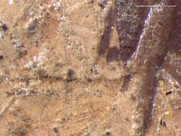
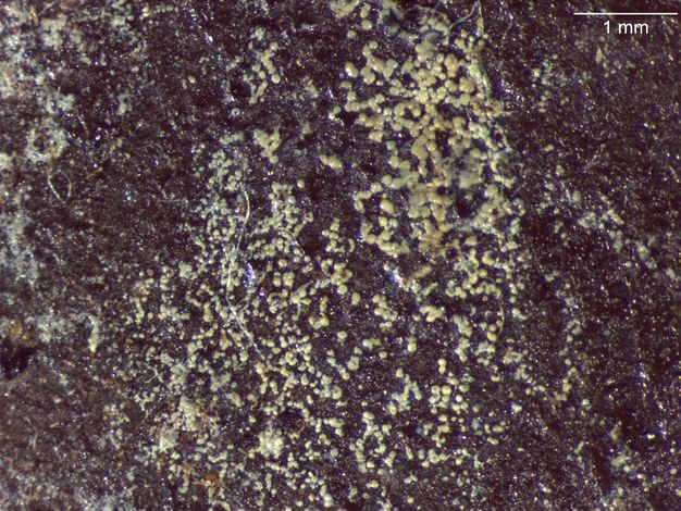
Irregular white hazy patches obscure the darker passages of the gray background toward the outer edges of the panel (fig. 14.7). The blanching is heavily concentrated along the background behind the sitter’s proper left, with some encrustations partially covering the left ear and hairline. Additional areas of blanching are present at the crown of the head and to the proper right of the hairpins. Under magnification, much of the blanching appears as a superficial crust comprised of fine, yellowish white grains of varying sizes (fig. 14.8).
The element chlorine is also readily mappable with this XRF apparatus and appears to explain the blanched appearance of parts of the painting where its distribution follows that of the lead at the surface (fig. 14.3G). High levels of chlorine are visible along the proper left edge of the panel, coinciding with the heaviest concentration of the crust in the background. A microsample of this white material from the surface of the proper left side of the black background, adjacent to the sitter’s hair, was further analyzed by SEM-EDX and found to consist of lead chloride without other detectable elements. This encrusted matter contained some beeswax medium identified by Fourier-transform infrared spectroscopy (FTIR) and had a gummy consistency. Unaltered lead pigment, when present in the sample, was distinguished by a finer particle size and lack of chloride. The implication of these white formations is that chloride salt, nearly ubiquitous in Egyptian artifacts from various sources and often derived from groundwater salinity or the burial environment, has interacted with the lead pigment to form lead chloride on the paint surface. Lead chloride is insoluble and thus favored to form in the interaction of most lead pigments with sodium chloride. This is a well-known phenomenon impacting not only wall paintings, but also easel paintings and lead-based architectural paints on masonry surfaces containing chlorides.14
A cross section from the forehead of a Tebtunis portrait (#6-21378b) in the PAHMA collection also contained a lead chloride compound as the main constituent in a white crust layer.15 A specific identification of the crust-forming compound as KPb2Cl5 was obtained in that case by synchrotron-based micro X-ray diffraction mapping of the cross section. Therefore, while the two cases are closely related, the end product is different.
Iron-Based Pigments Including Red Ochre
The map for iron shows the extensive use of iron earths in the artist’s palette (fig. 14.3H). Virtually every component of the composition relies upon iron in some form, including the red garment, all of the skin tones, and even the black in the hair. The overall red of the garment as well as its folds are rendered as variations in the distribution of this element. The rounding of the facial features in the later stages of painting that contrasts with their angularity in the Egyptian blue underdrawing is exemplified by their shapes in the iron map. The iron map also resolves pigment strokes in the hair more clearly than any other single element. It discloses the texture of the hair that could be interpreted as rows of braids at the front of the head, as well as a distinct boundary between the volume of the top of the head and that of the tightly braided hair bun in the back.
The depth of the tunic’s red color suggested that this area might make use of organic lake pigment in addition to the iron-based pigment found there with elemental mapping. Ultraviolet-induced visible fluorescence (UVF) imaging showed, however, no evidence of the distinctive pink/orange fluorescence of lake pigments, such as madder (fig. 14.9). The irregular, dull orange fluorescence visible with UVF in much of the red garment also extends into most areas of the portrait, with the exception of the face. A thin strip of the red tunic and striped collar next to the inner edge of the proper right clavus is also devoid of this orange fluorescence. It is more likely that this fluorescence corresponds to a clear surface coating that was not identified in this study. The tunic also appears dark in the false-color ultraviolet (FCUV) image, corroborating the presence of red ochre without the addition of a lake pigment (fig. 14.10).16
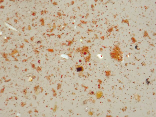
A microsample from the red tunic near the bottom edge of the panel was examined by polarized light microscopy (PLM) and SEM-EDX to determine whether the inorganic elements detected in the garment fully accounted for its red color. PLM shows that the sample only contains a dilute amount of very fine red iron oxide particles that have been well dispersed in an excess of wax medium, causing it to function like a colored varnish and deepening its color by reducing light scattering from the surface (fig. 14.11). It is not known if red lead has been incorporated into the painting’s composition elsewhere, but the lead map suggests a sparse presence of lead of unknown type in the upper regions of the red tunic (see figs. 14.3D and G). None was found in the PLM sample.
Restorations
A vertical split down the center of the panel shows evidence of both ancient and modern interventions. The paint has been consolidated in the worst areas of damage along the split in the head with a clear, glossy coating. UVF shows this coating has a milky, white fluorescence, but additional testing has not been carried out to identify it (see fig. 14.9). Due to the obvious presence of restoration coatings along the split, especially around the areas of paint loss in the head, it was assumed that repairs to the portrait occurred well after its completion, possibly at the time of excavation. However, no treatment documentation exists prior to or after its acquisition into the Nelson-Atkins collection.
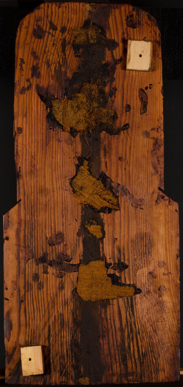
The X-radiograph revealed further information about repairs to the lower half of the split hidden below the surface. While the split becomes invisible on the surface below the mouth, a narrow strip of plain-weave fabric beneath the paint layers was identified in the X-radiograph. Its texture becomes visible at the mouth and extends to the bottom of the panel (see fig. 14.4). The red tunic exhibits cracking that follows the fabric texture in some spots, but further disruptions to the paint surface that could indicate this textile was applied as a later repair, such as reworking of the wax or inpainting, are absent from the lower half of the portrait. Other splits in the panel have not been bridged by fabric in this manner. The verso of the wood panel also has visible repairs along the crack. A dark brown resinous coating and sporadic patches of textile fragments are applied along the entirety of the vertical split (fig. 14.12). These textile fragments bear visual similarities to linen wrappings applied around funerary portraits when attached over the face of a mummy. This evidence suggests that the split in the panel was mended in preparation for painting.
Conclusions
From the time of its acquisition in 1937 until the undertaking of this technical study, the Nelson-Atkins’s Roman Egyptian funerary portrait had been interpreted exclusively by means of art historical inquiry. Analysis by XRF elemental mapping and complementary imaging techniques, augmented by microanalytical techniques when necessary, has revealed hitherto unknown aspects of material usage and artist’s practices. They reveal the use of Egyptian blue as an underdrawing, placing this painting within a larger corpus of funerary portraits that employed Egyptian blue as an additive to encaustic mixtures for flesh tones. The proportioning of carbon black and umber in the rendering of otherwise symmetrical dark passages of the painting reveal the painter’s attempt to render the illumination of the sitter. The painter’s choice of confining the use of lead white to the upper half of the portrait is an unusual one for which no definitive explanation exists. The ability to map the distribution of chlorine by XRF demonstrated a correlation of this element with highly visible haze formations on the dark background. Confirmation of the alteration of lead white to lead chloride in SEM-EDX of haze samples, and the lack of chloride in paint not similarly affected, strongly suggests that exposure of this lead pigment to environmental chlorides over the lifetime of the portrait is the underlying cause of blanching of the dark passages behind the figure’s head.
The analyses reported here have significantly furthered our understanding of the palette and working techniques in the Nelson-Atkins portrait. They have allowed us to explore both similarities and differences between this work and those investigated by other researchers, including some whose provenance is securely known. As a group, their working techniques are highly diverse and trends or groupings are only beginning to come into focus. Mineralogical analysis of the widely used earth pigments in the painting could advance our understanding of the individual pigments found in mixtures. The medium, which has not been investigated beyond FTIR confirmation that it is based upon beeswax, could provide further information about the painting through analysis by gas chromatography/mass spectrometry (GC/MS) methods. The wood and restoration materials remain yet to be identified as well. Future research of these materials will continue to broaden our knowledge of this portrait and its place within the history of Roman Egyptian funerary portraiture.
Acknowledgments
The authors would like to thank Kasey Hamilton for her contributions in imaging our portrait.
Notes
-
Doxiadis, Euphrosyne. 1995. The Mysterious Fayum Portraits: Faces from Ancient Egypt. Thames & Hudson.; Parlasca, Klaus. 1966. Mumienporträts und verwandte Denkmäler. Steiner.; Walker, Susan, and Morris Bierbrier, with Paul Roberts and John Taylor. 1997. Ancient Faces: Mummy Portraits from Roman Egypt. A Catalogue of Roman Portraits in the British Museum 4. The British Museum.. ↩︎
-
Walker, Susan, and Morris Bierbrier, with Paul Roberts and John Taylor. 1997. Ancient Faces: Mummy Portraits from Roman Egypt. A Catalogue of Roman Portraits in the British Museum 4. The British Museum.. ↩︎
-
Taggart, Ross E., ed. 1959. Handbook of the Collections in the William Rockhill Nelson Gallery of Art and Mary Atkins Museum of Fine Arts. 4th ed. William Rockhill Nelson Gallery of Art and Mary Atkins Museum of Fine Arts.; Vermeule, Cornelius. 1964. “Greek and Roman Portraits in American Collections Open to the Public: A Survey of Important Monumental Likenesses in Marble and Bronze Which Have Not Been Published Extensively.” Proceedings of the American Philosophical Society 108 (2): 99–134.; Doxiadis, Euphrosyne. 1995. The Mysterious Fayum Portraits: Faces from Ancient Egypt. Thames & Hudson.. ↩︎
-
Dyer, Joanne, and Nicola Newman. 2020. “Multispectral Imaging Techniques Applied to the Study of Romano‑Egyptian Funerary Portraits at the British Museum.” In Svoboda and Cartwright 2020.. ↩︎
-
De Viguerie, Laurence, Sophie Rochut, Matthias Alfeld, Philippe Walter, Sophie Astier, Valérie Gontero, and Florence Boulc’h. 2018. “XRF and Reflectance Hyperspectral Imaging on a 15th-Century Illuminated Manuscript: Combining Imaging and Quantitative Analysis to Understand the Artist’s Technique.” Heritage Science 6: 11. https://doi.org/10.1186/s40494-018-0177-2.. ↩︎
-
Solé, V. Armando, Emmanuel Papillon, Marine Cotte, Philippe Walter, and Jean Susini. 2007. “A Multiplatform Code for the Analysis of Energy-Dispersive X-Ray Fluorescence Spectra.” Spectrochimica Acta Part B: Atomic Spectroscopy 62 (1): 63–68.. ↩︎
-
Verri, Giovanni. 2009. “The Application of Visible-Induced Luminescence Imaging to the Examination of Museum Objects.” Proceedings SPIE 7391, O3A: Optics for Arts, Architecture, and Archaeology 2: 739105–12. https://doi.org/10.1117/12.827331.; Accorsi, Gianluca, Giovanni Verri, Margherita Bolognesi, Nicola Armaroli, Catia Clementi, Costanza Miliani, and Aldo Romani. 2009. “The Exceptional Near-Infrared Luminescence Properties of Cuprorivaite (Egyptian Blue).” Chemical Communications, no. 23, 3392–94. https://doi.org/10.1039/B902563D.. ↩︎
-
See Mahon et al., this volume; Mayberger, Evelyn (Eve), Jessica Arista, Marie Svoboda, and Molly Gleeson. 2020. “Invisible Brushstrokes Revealed: Technical Imaging and Research of Romano-Egyptian Mummy Portraits.” In Svoboda and Cartwright 2020.. ↩︎
-
Ganio, Monica, Johanna Salvant, Jane Williams, Lynn Lee, Oliver Cossairt, and Marc Walton. 2015. “Investigating the Use of Egyptian Blue in Roman Egyptian Portraits and Panels from Tebtunis, Egypt.” Applied Physics A 121 (3): 813–21. https://doi.org/10.1007/s00339-015-9424-5.. ↩︎
-
Thiboutot, Gabrielle. 2020. “Egyptian Blue in Romano-Egyptian Mummy Portraits.” In Svoboda and Cartwright 2020.. ↩︎
-
Dyer, Joanne, and Nicola Newman. 2020. “Multispectral Imaging Techniques Applied to the Study of Romano‑Egyptian Funerary Portraits at the British Museum.” In Svoboda and Cartwright 2020.; Park, Hae-Min, Amaris Sturm, Glenn Gates, and Lisa Anderson-Zhu. 2019. “Findings from an Examination of Two Mummy Portraits.” The Journal of the Walters Art Museum 74. https://journal.thewalters.org/volume/74/note/the-walters-portrait-mummies/.; Sabino, Rachel C., Ken Sutherland, Emeline Pouyet, Federica Pozzi, and Marc Walton. 2019. “Surprise Encounters with Mummy Portraits at the Art Institute of Chicago.” AIC Paintings Specialty Group Postprints 31.. ↩︎
-
Thiboutot, Gabrielle. 2020. “Egyptian Blue in Romano-Egyptian Mummy Portraits.” In Svoboda and Cartwright 2020.. ↩︎
-
Park, Hae-Min, Amaris Sturm, Glenn Gates, and Lisa Anderson-Zhu. 2019. “Findings from an Examination of Two Mummy Portraits.” The Journal of the Walters Art Museum 74. https://journal.thewalters.org/volume/74/note/the-walters-portrait-mummies/.. ↩︎
-
Ordonez, Eugena, and John Twilley. 1998. “Clarifying the Haze: Efflorescence on Works of Art.” Analytical Chemistry 69 (13): 416A–22A. Reprinted without color illustrations in WAAC Newsletter 20 (1). https://cool.culturalheritage.org/waac/wn/wn20/wn20-1/wn20-108.html.. ↩︎
-
Salvant, Johanna, Jane Williams, Monica Ganio, Francesca Casidio, Céline Daher, Ken Sutherland, Letizia Monico, Frederik Vanmeert, Steven de Meyer, Koen Janssens, Caroline R. Cartwright, and Marc Walton. 2018. “A Roman Egyptian Painting Workshop: Technical Investigation of the Portraits from Tebtunis, Egypt.” Archaeometry 60 (4): 815–33. https://doi.org/10.1111/arcm.12351.. ↩︎
-
Dyer, Joanne, and Nicola Newman. 2020. “Multispectral Imaging Techniques Applied to the Study of Romano‑Egyptian Funerary Portraits at the British Museum.” In Svoboda and Cartwright 2020.. ↩︎