Introduction
The study of Egyptian mummy portraits has become an appealing and enlightening topic. These funerary portraits are not only timeless visual documents that represent the individuals who lived in Egypt between the first and third centuries CE but also valuable resources in the development and understanding of ancient painting technology. Archaeologists such as Sir William Flinders Petrie, Albert Gayet, papyrologists Bernard P. Grenfell and Arthur S. Hunt, and the prominent Viennese art dealer Theodor Graf brought many of these distinctive and evocative artifacts to light in the nineteenth century.1 Since their discovery, most of these painted panels have been housed in museums and private collections worldwide, detached from their original contexts: their mummified bodies. This group of ancient painted artifacts also includes funerary panels depicting individuals and deities, some with their original frames or pintles still intact.
In all, these specialized works provide an important connection to the artisans, their materials, methods, innovations, and clues for the transfer of ideas in antiquity. The excellent state of preservation of many of these works affords an unprecedented opportunity for research, increasing our understanding of ancient pigments, binding media, wood, textiles, and coatings. Throughout their histories, however, the intervention of many detached paintings involved consolidation, inpainting, flattening, backing, coatings, and alteration by combining different panels, both ancient and modern,2 raising questions in the study of the materials and techniques used in their production. Fortunately, not all portraits have been disturbed and funerary paintings continue to be discovered, shedding new light on artistic practice and ancient trade in Roman Egypt.3
Unique observations and features of these paintings have come to light through the collaborative model of the J. Paul Getty’s APPEAR project, which fosters examination and evaluation of funerary portraits using interdisciplinary scholarship, comparative studies, and scientific analyses.4 One such observation, and the focus of this paper, was that of a distinct surface coating identified on a group of funerary panel paintings housed in five separate museum collections. A subsequent project, which evolved to investigate this unique feature, brought together a multidisciplinary team of antiquity conservators, conservation scientists with expertise in organic material studies, and a researcher specializing in radiocarbon dating (14C).
Funerary Portraits
Funerary mummy portraits are images of deceased individuals painted on a wide variety of wooden panels or textile shrouds that were secured over the faces of their mummified bodies. Other types of funerary panel paintings, not attached to mummies, were likely used in private homes, possibly in a domestic shrine context, although their original functions are unknown.5 In this paper the terms portrait and funerary portrait are both used to describe funerary and mummy paintings. These painted artifacts, produced exclusively in Egypt,6 were adopted from the Greco-Roman painting tradition and incorporated into the Egyptian funerary practice. The paintings, which represented both personalized and divine images, were reserved only for those of a “certain social group.”7 Although the pharaonic tradition of preserving human remains spanned over five thousand years, the later practice of creating personalized portraits lasted only about four hundred years, likely because of shifting external influences and cultural diversity within Egypt. By the late fourth century CE, after the edict of Theodosius banned pagan rituals in Egypt ca. 379 CE, mummification practices were being phased out.8
Ancient funerary paintings were created on textile shrouds or on panels of a variety of local and imported woods. Both supports were specially prepared for their unique purpose. They were painted using an impressive palette of natural or manufactured pigments bound with organic-based media. Today, a renewed interest in ancient paintings, together with the application of sensitive and specific analytical techniques and access to multidisciplinary corroborative studies, has broadened our understanding of ancient painting technology, enabling new discoveries.9 For example, studies of twenty-two water-based binding media (tempera) portraits using gas chromatography/mass spectrometry (GC/MS) and enzyme-linked immunosorbent assay (ELISA) have established that animal glue was the predominant medium used in antiquity (90%), followed by plant gums (10%).10 Likewise, analyses of encaustic binding media have confirmed that their composition can involve more than just pure beeswax.11
Preliminary Investigations
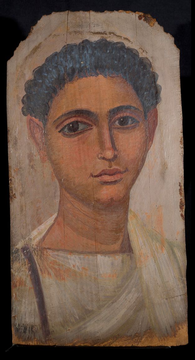
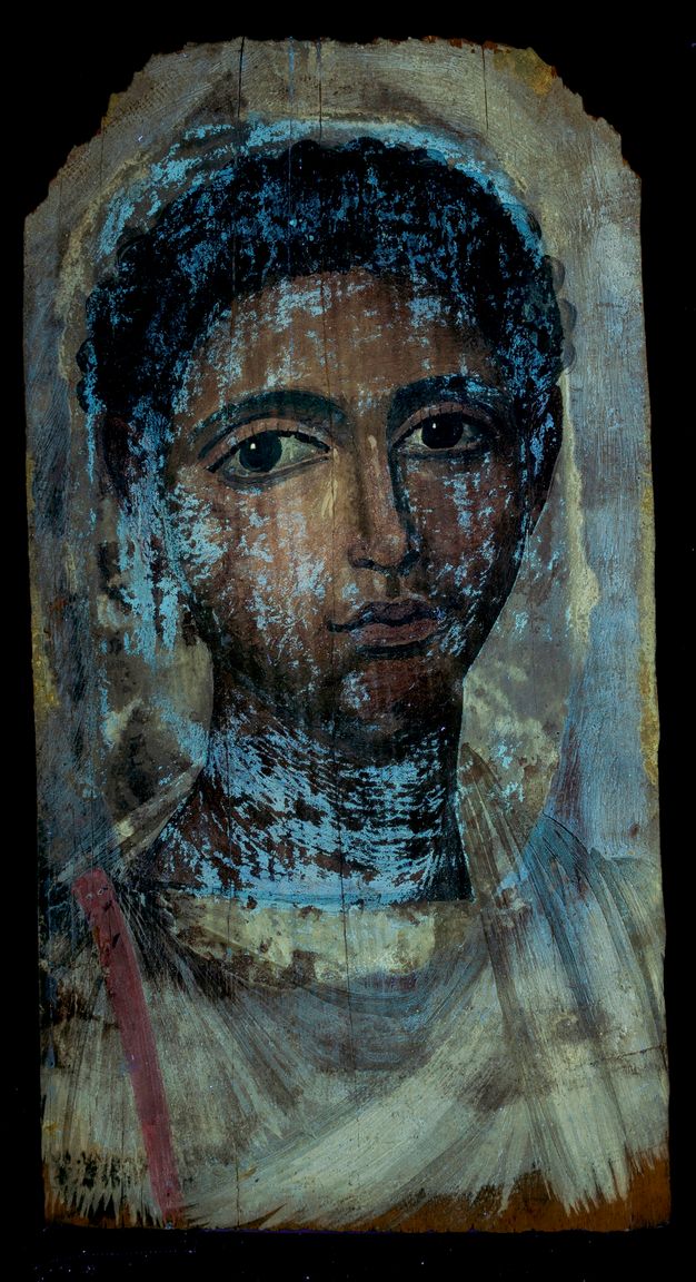
In 2008, a study at the J. Paul Getty Museum using ELISA confirmed the presence of an animal glue binder on the tempera funerary Portrait of a Bearded Man (74.AP.20; fig. 8.1).12 The study also detected traces of egg in the paint, raising the question of whether it was a component of the ancient binding media or a modern conservation intervention. Subsequent analyses in 2014 using GC/MS in tandem with ultraviolet fluorescence microscopy corroborated the presence of egg on that funerary portrait as well as on two other Getty panel paintings (74.AP.21 and 74.AP.22) once associated with the bearded man portrait.13 Further research revealed that the egg was present as an isolated surface layer, but at that time it was noted that it “may be an artifact due to modern restoration of the portraits.”14
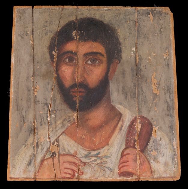
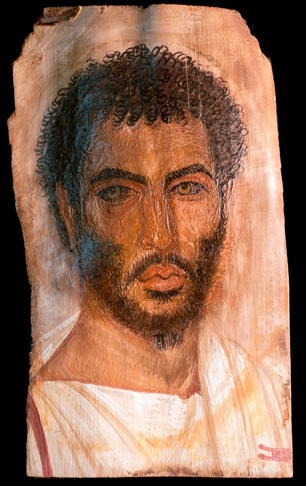
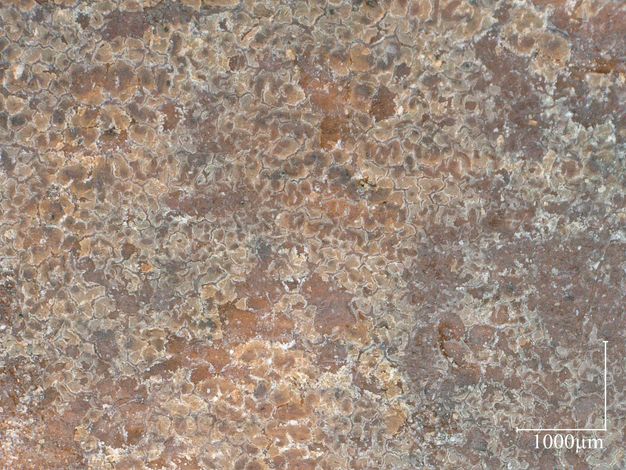
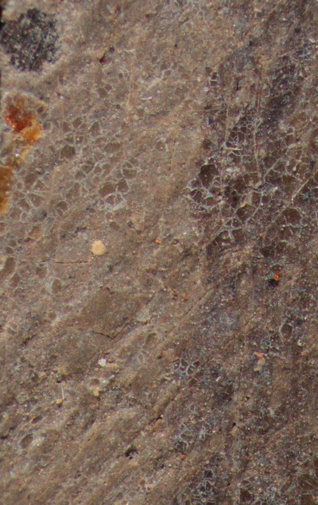
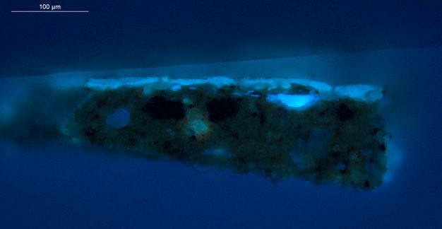
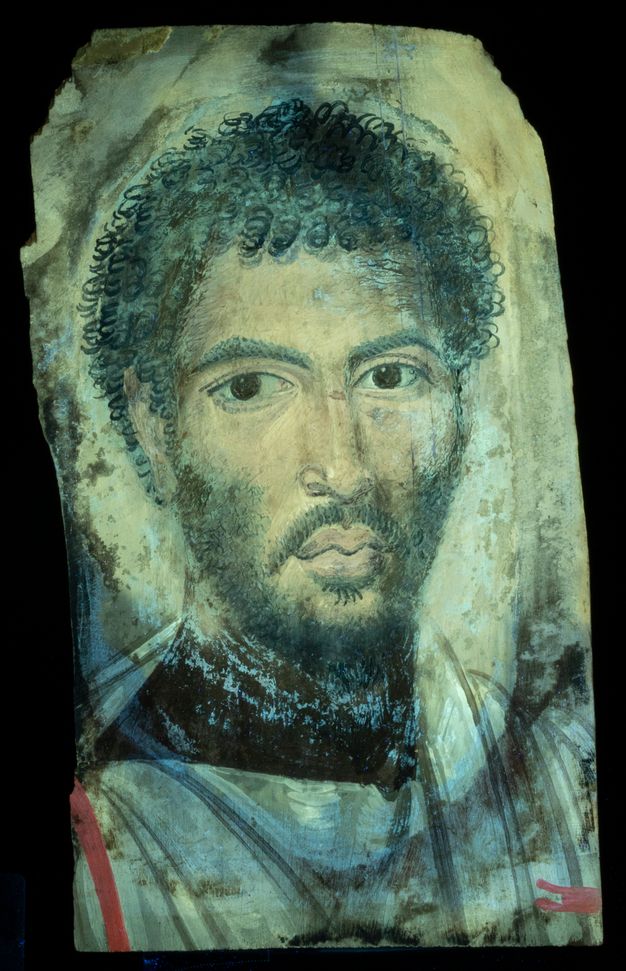
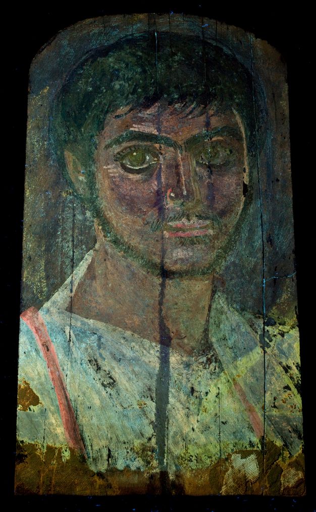
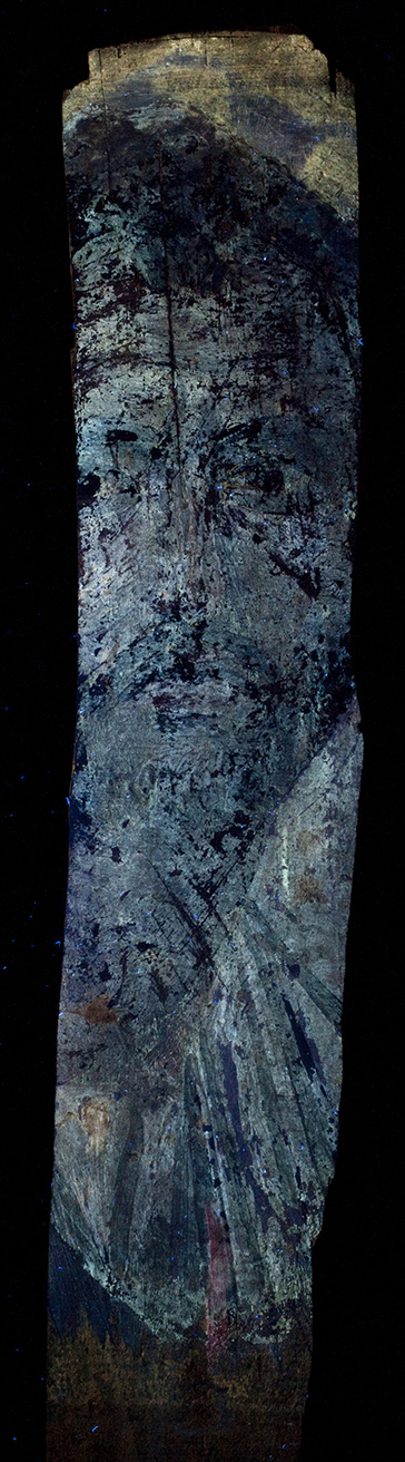
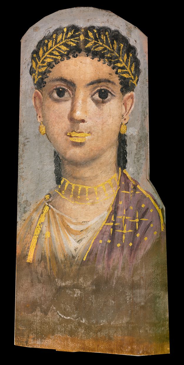
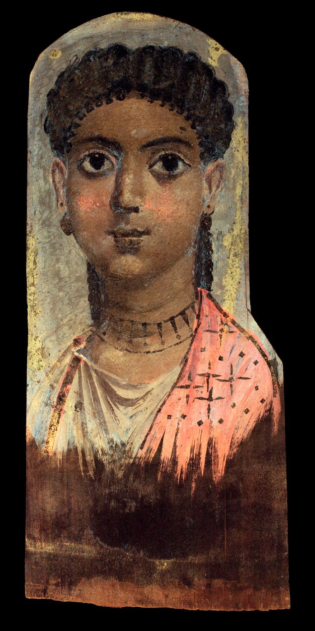
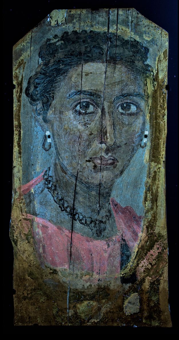
At the first APPEAR project meeting in 2013, participants from a variety of museums discussed the presence of an unusual surface deposit on the mummy portrait of a bearded man in the Getty’s collection (74.AP.11) as a possible future research topic (figs. 8.2 and 8.3). The surface feature was described as an “accretion or scum,” possibly related to the encaustic medium or the production of lead soaps.15 This open discussion led to the identification of a similar surface feature on two encaustic portraits in Copenhagen’s Ny Carlsberg Glyptotek collection (ÆIN 681 and 684; figs. 8.4A–8.5B). GC/MS analysis of the surface accretion on the Glyptotek and Getty mummy portraits confirmed the presence of egg.16 Observation of a paint cross section from Glyptotek ÆIN 684 with ultraviolet fluorescence microscopy corroborated that the egg was present as a surface coating (fig. 8.6).17 Unlike the media commonly used to execute funerary panel paintings, such as beeswax, oil, and pine resin, or water-based media such as animal glue and plant gums, this surface coating could be visualized on the three portraits with ultraviolet radiation as an uneven, bluish fluorescence (figs. 8.7–8.9). The visible fluorescence extended only to where the linen wrappings would have covered the panel, securing it to its mummy. A further diagnostic feature could be observed with magnification: the coating appeared as yellowed “islands” of a brittle surface encrustation (see figs. 8.3, 8.4B, and 8.5B). The presence of egg was unexpected since previous analyses had shown that the paint medium of three of these four portraits was encaustic and that the medium of the other was animal glue, leaving the significance and source of the egg unclear. These initial portrait analyses provided a starting point for in-depth analysis of the egg coatings by peptide mass fingerprinting (PMF) and liquid chromatography with tandem mass spectrometry (LC-MS/MS).


Methods: PMF and LC-MS/MS
Advanced proteomics techniques such as PMF and LC-MS/MS, which have been adopted and adapted from biotech into cultural heritage, can provide precise identification of proteinaceous media and coatings and even determine the species of origin of those materials.18 PMF analysis involves the enzymatic digestion of proteins followed by Matrix Assisted Laser Desorption-Ionization Time of Flight Mass Spectrometric (MALDI) analysis of the resultant peptide mixture to produce a “peptide mass fingerprint.”19 Marker ions in the MALDI spectra from known reference materials are compared with those from unknown samples for identification.20 LC-MS/MS with database searching is a mainstay of biotechnology. Its sensitivity is ideally matched to the minimal samples generally available from cultural objects, and its specificity allows more precise identification of proteinaceous materials than may be possible with other techniques.21 In the present work, LC-MS/MS was used to identify proteinaceous materials or to confirm those found with PMF.
The PMF and LC-MS/MS experimental details have been published previously and are briefly described here.22
Samples
Samples from the Glyptotek portraits were remnants from previous analyses.23 All other samples were obtained by conservators using sample sticks, which are polystyrene strips with adhered fiber-optic polishing film to abrade and entrap a small amount of material for analysis (fig. 8.10). All portraits had a clearly visible surface layer and characteristic visual appearance, and the UVF response guided the conservators to specific sample sites.

Digestion
60 μL of 50 mM ammonium bicarbonate (AMBI) was added to each sample in a 600 μL Eppendorf tube and heated to 75°C for 60 min. After cooling, 8 μL Promega Sequence Grade trypsin (0.02 μg/μL in 50 mM AMBI) was added and digestion proceeded overnight at 37°C.
MALDI
2 μL of the digest were added to 20 μL 40% acetonitrile (ACN), 0.1% trifluoroacetic acid with saturated α-Cyano-4-hydroxycinnamic acid matrix; 0.65 μL of the mixture was spotted onto the MALDI plate. Spectra were obtained with an Applied Biosystems/Sciex 5800 MALDI-TOF/TOF instrument operated in positive reflector mode. Acquired spectra were exported from Applied Biosystems Data Explorer software as text files and imported into mMass,24 where spectra were manually inspected for markers.
LC-MS/MS
Thermo Q Exactive-Orbitrap interfaced with an UltiMate™3000 chromatographic system. The mass spectrometer was operated in data-dependent mode (full scan 350–1800 Da, resolution 70,000; top 10 ions were selected for fragmentation, resolution 17,500; detected ions were placed on an exclusion list for 10 sec.). LC buffers: A: 0.1% formic acid, B: 0.1% formic acid in ACN; gradient from 2–50% B over 60 min. One microliter of each of the digests analyzed by PMF was injected onto a nanocolumn: 75 μm ID x 15 cm long, packed with Reprosil AQ C18 with 1.9 μm beads (Dr. Maisch, Germany). LC-MS/MS data files were converted to mascot generic format (MGF), Matrix Science,25 with msConvert software from ProteoWizard26 and searched online through Mascot.
Results
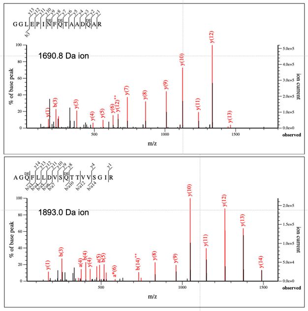
PMF sample coating analysis on two Getty and two Glyptotek portraits (see figs. 8.1, 8.2, 8.4A, 8.5A) corroborated the previous findings of egg and, in addition, found that it is whole hen egg (yolk and white together). Figure 8.11 compares MALDI spectra for two of the portraits together with a spectrum from whole hen egg. Several of the more intense egg-related ions are labeled. Unlabeled ions are mainly contaminating keratin. The insets are examples illustrating that isotopic clusters for peptides with asparagine (N) and/or glutamine (Q) residues in the portrait spectra are shifted to higher mass due to a high degree of deamidation in the samples.27 LC-MS/MS analysis of the same digests confirmed the PMF conclusions identifying proteins indicative of hen egg yolk and glair that is highly deamidated. Although the extent of deamidation can be influenced by a variety of factors, in the authors’ experience it seems excessive, raising the possibility that the high degree of deamidation is the result of the preparation or application of the coating. Figure 8.12 shows the MS/MS spectra for the ions from Getty 74.AP.11, illustrated in the insets in figure 8.11. Based on these results, future analyses can be confidently done with PMF alone.

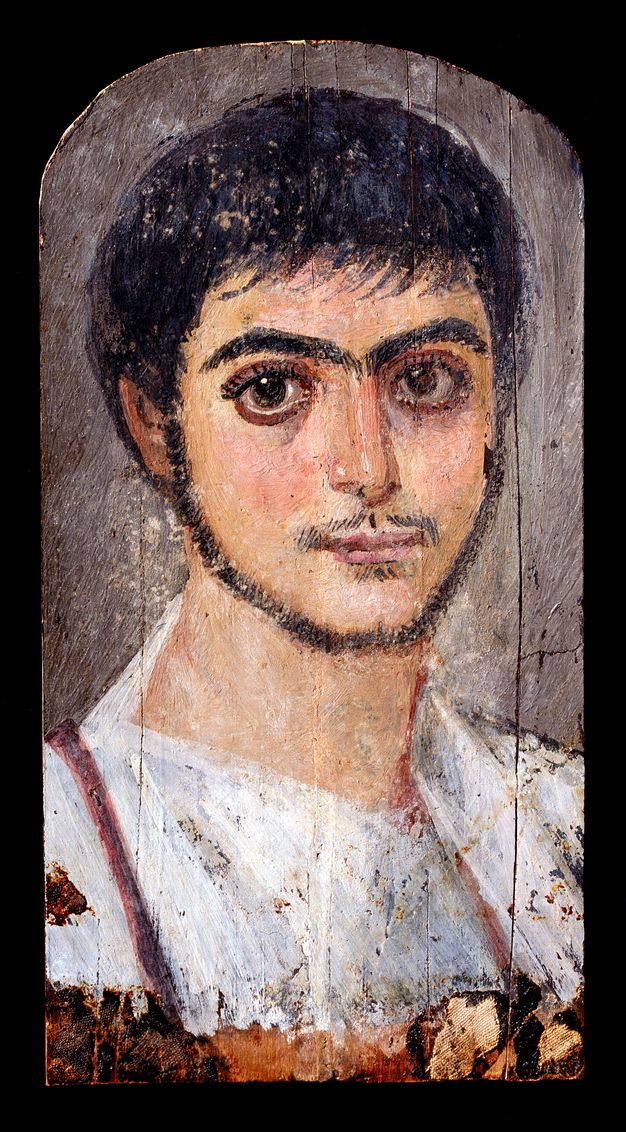
Following confirmation of the presence of whole hen egg on the four panels, the APPEAR database was searched for UV images of other portraits exhibiting the characteristic fluorescence.28 Three other panels were identified based on the observed diagnostic features, one each from the collections at the Norton Simon Museum, Pasadena (F.1978.19.P), the Cleveland Museum of Art (1971.137), and the Kunsthistorisches Museum in Vienna (X 297) (figs. 8.13A–8.15B). These institutions agreed to take microsamples of the surface coating using sample sticks, as illustrated in figure 8.10, and submit them for analysis.29 Both PMF and LC-MS/MS confirmed the presence of deamidated whole hen egg on the Norton Simon Museum and the Kunsthistorisches Museum portraits, and deamidated hen egg glair on the portrait from the Cleveland Museum of Art. Results for the seven analyzed portraits can be found in figure 8.16.
| Institution | Accession No. | Medium | ELISA | GCMS | PMF Result | LCMSMS Result |
|---|---|---|---|---|---|---|
| J. Paul Getty | 74.AP.11 | Encaustic | Egg | Egg | Whole hen egg | Whole hen egg, mammalian collagen |
| J. Paul Getty | 74.AP.20 | Tempera | Egg, glue | Glue | Whole hen egg, trace collagen | Whole hen egg, mammalian collagen |
| Glyptotek | ÆIN 681 | Encaustic | N/A | Egg | Whole hen egg | Whole hen egg, mammalian collagen |
| Glyptotek | ÆIN 684 | Encaustic | N/A | Egg | Whole hen egg | Whole hen egg, mammalian collagen |
| Norton Simon | F.1978.19.P | Encaustic | N/A | N/A | Whole hen egg | Whole hen egg, mammalian collagen |
| Vienna | X 297 | Encaustic | N/A | N/A | Whole hen egg | Whole hen egg, mammalian collagen |
| Cleveland | 1971.137 | Encaustic | N/A | N/A | Hen egg glair | Hen egg glair, mammalian collagen |
Hypothesis
To address why the observed coating does not extend to the edges of the mummy portrait panels, we initially hypothesized that it was applied after their discovery in the late nineteenth century to protect the nearly two-thousand-year-old painted surface. This implies that the coating may have been added before the panel was removed from its mummified remains, possibly to saturate or consolidate the surface. It is probable that, when recently excavated portrait mummies reached the hands of collectors and art dealers with a desire to improve (revive or treat) their appearance, a coating may have been applied. The use of egg white (glair) as a “varnish” on paintings was a common practice in the nineteenth century.30 This theory conveniently explains the egg identified on the funerary panel of a bearded man (see fig. 8.1), which had not been attached to a mummy. Another hypothesis was that this coating had been applied in antiquity, after the panels were inserted into the mummy wrappings. While applying libations as part of the funerary ritual was a well-known Pharaonic practice, the application of ritual coatings is not known on Roman Egyptian portrait mummies.31
Radiocarbon Dating
Radiocarbon dating (14C) was used to determine when the coatings were applied. Ancient organic materials dating is possible by measuring carbon-14 (14C) isotopes that are absorbed, via photosynthesis or consumption, by living organisms. Based on this, it is possible to determine an approximate date when an organism had died through the measurement of 14C decomposition. In 2020, the W.M. Keck Carbon Cycle Accelerator Mass Spectrometer facility at UC Irvine was contacted about the possibility of dating the egg coating.32 This facility had dated milligram-sized samples of wood and textile from eleven mummy portraits in the Getty collection in 2013, and it was now possible to obtain age data from micro samples of egg, weighing approximately one tenth of a milligram.
Two portraits were selected: the Getty’s Mummy Portrait of a Bearded Man (74.AP.11) and the Norton Simon’s Portrait of a Man (F.1978.19.P). Microsamples (approx. 0.25 mgC) were collected from the surface of each panel under magnification using a scalpel. The 14C results for the whole hen egg on the Getty portrait indicates that it was applied around 92 CE (median probability). This date is consistent with the age range of the portrait’s linden wood panel, analyzed at the same facility, and corresponding to a median probability calibrated date of 103 CE.33 The median probability calibrated radiocarbon date for the egg coating on the Norton Simon portrait was determined to be 24 CE.
The dates acquired for both portraits indicate that the egg coatings were applied in antiquity and are not modern additions (fig. 8.17). Although the coatings on only two of the portraits in this study have an established date of application, it is probable that the other portraits showing similar diagnostic features were also applied in antiquity. This theory is further corroborated by the confirmation of highly deamidated hen egg as established by PMF and LC-MS/MS. Although not comprehensive, the evidence of even two ancient dates for the egg coating provides new insights to artistic and/or funerary practices in Egypt between the first and second centuries CE.
| Institution | Accession No. | Medium | Calibrated 14C Date (95% confidence interval) / Median probability |
|---|---|---|---|
| J. Paul Getty | 74.AP.11 | Encaustic | 25–128 CE / 92 CE |
| J. Paul Getty | 74.AP.20 | Tempera | |
| Ny Glyptotek | ÆIN 681 | Encaustic | |
| Ny Glyptotek | ÆIN 684 | Encaustic | |
| Norton Simon | F.1978.19.P | Encaustic | 42 BCE–108 CE / 24 CE |
| Kunsthistorisches, Vienna | X 297 | Encaustic | |
| Cleveland | 1971.137 | Encaustic |
The egg detected is specifically from Gallus gallus domesticus, the domesticated chicken, exploited in antiquity for sport, prophesy, and, during Egypt’s Ptolemaic period, as an important food source. Analysis of the egg coating by PMF and LC-MS/MS indicated that it was highly deamidated, although it is uncertain whether deamidation here is related to age, burial conditions, or another type of “alteration,” possibly in preparing the egg for application.
The Chicken (Gallus gallus domesticus)
Although archaeological evidence for the domesticated chicken is rare and their origin is still a matter of debate, it is generally believed that the chicken originated in Southeast Asia and made its way westward into Mesopotamia around 2000 BCE.34 Shortly after the chicken’s arrival in Egypt, the rooster gained popularity as a source of entertainment (cockfighting), a sport also popular in Greece and Rome.35 The Romans also revered the rooster for its ability to predict the success or failure of battles.36 As a result, roosters were viewed as sacred and protected, typically living long lives as a result.37 In Egypt, following a mysterious period of disappearance, the chicken returned during the Ptolemaic period around 305 BCE, when unique and sophisticated egg incubators were invented, prompting the development of an industry that made chicken eggs widely available.38
The ancient Egyptians documented the animals and birds they knew and used. Illustrated in hunt and banquet scenes, the more exotic species such as partridges, pigeons, ducks, geese, quail, herons, ibis, and hawks were commonly depicted, while the chicken was rarely included in the artistic repertoire.39
History and Use of “Varnish” Coatings
In the early nineteenth century, the chemist Alfred Lucas was the first to identify and study the composition of coatings and varnishes (clear, pigmented, and opaque) applied on Egyptian artifacts between the first half of the Eighteenth and the Twenty-Sixth Dynasties (ca. 1549–525 BCE).40 While Lucas’s work was pivotal, in-depth investigations of these coatings today have enabled more precise identifications, expanding our understanding of their source and function.41 Believed to have been used primarily for ritual purposes, various materials such as bitumen, pistache, and pine resins (sntr-pistacia and pinus) have consistently been identified on painted funerary material.42
While egg varnishes applied to artworks were common from the fourteenth through nineteenth centuries CE,43 the reliable identification of egg as a coating in ancient Egypt has not been documented to date.44 In his study of ancient Egyptian materials, Lucas discussed the identification of egg (albumin) possibly used as an ancient binder or adhesive, but states that results could not be verified.45 Such complex investigations have significantly advanced through the development of more sensitive analytical instrumentation and techniques that enable the precise identification of aged materials.46 Yet the question remains, why were the Roman Egyptian funerary panels coated with hen egg?
Artistic Varnishes
While various types of varnishes and glazes identified on painted artifacts were likely applied as part of religious ceremonies, their aesthetic function is also highly probable. Selectively applying a coating, sometimes pigmented, to create different striking visual impressions such as a contrasting matte and shiny surface is often described.47 For example, coatings from a Nineteenth Dynasty (ca. 1298–1187 BCE) wall painting in the tomb of Nefertari and from Twenty-Second Dynasty (ca. 948–743 BCE) coffins contained pigmented pistacia resin to create a contrasting shiny/matte and golden-colored appearance.48 While both natural tree resin and egg white were detected on the Nefertari wall paintings, however, traces of egg albumen could not be confirmed as ancient due to the extensive amount of modern restoration.49
Thus far, evidence for the use of egg has not been firmly identified for rituals or as protective coatings in the funerary context in Egypt during the first to third centuries CE. Therefore, evidence presented here of a hen egg coating, confirmed as ancient on two Roman-Egyptian painted panels, reveals a unique function, potentially introducing a new practice to an enduring Egyptian funerary ritual.50
Egg Symbolism
The powerful symbolism of the egg in antiquity cannot be overlooked. Its significance was recognized in Hellenic cultures as an offering and as a symbol of rebirth.51 The egg also has deep resonance as part of the Egyptian creation myth, representing rebirth and the renewal of life.52 As a cosmic motif, the egg is connected to the continuation of human breath after death.53 The egg embodies divinities, gods of creation such as Thoth and Amun, Isis and Osiris, and their son Horus, born as the sun god in the form of an egg.54 It should not be surprising that the deeply symbolic egg would be selected as a material for anointing the deceased, to protect and promote rebirth in the afterlife. Its ubiquitous significance emphasizes the multicultural influences intertwined with ritual practices that make the production of portrait mummies so unique.
This study required the collaboration of a multidisciplinary team, each participant making very important contributions to the investigation. The ability to identify parallel features and acquire samples through collaborative projects, such as the APPEAR database, has enabled this study to evolve. Furthermore, characterizing these discrete materials using various investigative techniques, such as imaging, microscopic examination, and organic analysis, has established the thread that defines a unique group of artifacts, potentially revealing a new and unique practice. The ability to confirm the egg source on seven funerary paintings using highly sensitive analytical techniques, as well as the confirmation of an ancient date by 14C for the application of the coating on two mummy portraits, presents an important direction of research fostering continued corroboration.
GC/MS and/or ELISA analyses of paint collected from the Getty and Glyptotek portraits, three of which were painted with encaustic and one funerary panel with tempera, initiated the investigation into the presence of egg. This led to the identification and analyses of three other portraits from the APPEAR database with similar diagnostic features. Identifying egg in the encaustic portraits is significant as it was unexpected, resulting in the discovery that egg was not a component of the binder but was, in fact, a superficial coating. Localized investigation of the egg coating by PMF and LC-MS/MS analyses confirmed that it was whole hen egg in six of the seven portraits analyzed within this group; egg white was detected on the seventh. Furthermore, the egg on all seven was deamidated, suggesting they had undergone a type of alteration, a potential direction for future study. 14C dating of two samples from the Getty (74.AP.11) and Norton Simon (F.1978.19.P) mummy portraits confirmed that the egg coatings were ancient and not modern interventions as was originally believed.
Although the ancient 14C date was confirmed on only two portraits, we can deduce that, based on the identification of deamidated hen egg on seven painted panels, corroborated by their visual appearance and method of application, this may have been an Egyptian funerary practice. However, the reason for its application remains inconclusive. Was it a method to protect and saturate the surface? Was it an artistic effort to enhance the surface sheen and reflectivity, or was it a ritual practice symbolizing creation and renewal in the afterlife? These questions will remain unanswered until more ancient panel paintings are identified and studied. Foremost, this research stresses the need for more comparative studies and emphasizes the value of collaborative research to better understand ancient materials and methodology and their function.
Acknowledgments
The authors would like to thank the institutions that generously allowed sampling of their portraits to make this study possible: the Norton Simon Museum; the Cleveland Museum of Art; Kunsthistorisches Museum, Vienna; Ny Carlsberg Glyptotek; and the J. Paul Getty Museum. MS graciously thanks Richard Newman, Kathlyn Cooney, Salima Ikram, Alessia Amenta, and Tamar Hodos for sharing their valuable knowledge of egg, and Susanne Gänsicke and Yosi Pozeilov for their support; DK thanks Sue Abbatiello, formerly of Northeastern University, for sharing her expertise in LC-MS/MS; JM thanks Michael Schilling; LRS is grateful to the Carlsberg Foundation, the Getty Conservation Institute, and the staff at the National Museum of Denmark and the Ny Carlsberg Glyptotek for their kind permission to examine and take samples from their mummy portraits.
Notes
-
Barr, Judith. 2020. “From All Sides: The APPEAR Project and Mummy Portrait Provenance.” In Svoboda and Cartwright 2020.; Bierbrier, Morris. 1997. “The Discovery of the Mummy Portraits.” In Walker and Bierbrier 1997.. ↩︎
-
Jaeschke, Richard L. 1997. “Mechanical Cleaning and the Conservation of Portraits from the Petrie Museum of Egyptian Archaeology.” In Portraits and Masks: Burial Customs in Roman Egypt, edited by Morris Bierbrier. The British Museum.. ↩︎
-
Gehad, Basem, Lorelai H. Corcoran, Mahmoud Ibrahim, Ahmed Hammad, Mohamed Samah, Abd Allah Abdo, and Omar Fekry. 2022. “Newly Discovered Mummy Portraits from the Necropolis of Ancient Philadelphia–Fayum.” BIFAO 122, 245–64. https://journals.openedition.org/bifao/11727.. ↩︎
-
Svoboda, Marie, and Caroline R. Cartwright, eds. 2020. Mummy Portraits of Roman Egypt: Emerging Research from the APPEAR Project. J. Paul Getty Museum.. ↩︎
-
Borromeo, Georgina E., Ingrid A. Neuman, Scott Collins, Catherine Cooper, Derek Merck, and David Murray. 2020. “Framing the Heron Panel: Iconographic and Technical Comparanda.” In Svoboda and Cartwright 2020.; Stevens, Anna. 2009. “Domestic Religious Practices.” In UCLA Encyclopedia of Egyptology, edited by Willeke Wendrich and Jacco Dieleman. University of California, Los Angeles. http://escholarship.org/uc/item/7s07628w.. ↩︎
-
Anita Kirchhof and A. Rehorovics, “A Face from the Past . . . Painted Portrait on Wood from a Mummy Grave in Aquincum,” in Bragantini, Irene. 2010. Atti del X Congresso internazionale dell’AIPMA, Association internationale pour la peinture murale antique, Napoli 17-21 Settembre 2007 / a cura di Irene Bragantini. Annali di Archeologia e Storia Antica. Quaderni / Dipartimento di Studi del Mondo Classico e del Mediterraneo Antico, no. 18. Napoli: Universitá degli Studi di Napoli “L’Orientale.”. ↩︎
-
Walker, Susan. 1997. “Mummy Portraits in Their Roman Context.” In Portraits and Masks: Burial Customs in Roman Egypt, edited by Morris L. Bierbrier. The British Museum.. ↩︎
-
Pharr, Clyde, Theresa Sherrer Davidson, and Mary Brown Pharr, trans. 2001. The Theodosian Code and Novels, and the Sirmondian Constitutions. The Lawbook Exchange.. ↩︎
-
Verri, Giovanni, Thorsten Opper, and Thibaut Devièse. 2010. “The ‘Treu Head’: A Case Study in Roman Sculptural Polychromy.” The British Museum Technical Research Bulletin 4: 39–52.; Verri, Giovanni. 2014. “Roman Sculpture and Colour: The ‘Treu Head’” (Ancient Greek and Roman Color/Polychromy). Video, 9 min., 6 sec. https://www.youtube.com/watch?v=gRMPYh2QdSM.; Svoboda, Marie, and Caroline R. Cartwright, eds. 2020. Mummy Portraits of Roman Egypt: Emerging Research from the APPEAR Project. J. Paul Getty Museum.. ↩︎
-
Mazurek, Joy, Marie Svoboda, and Michael Schilling. 2019. “GC/MS Characterization of Beeswax, Protein, Gum, Resin, and Oil in Romano-Egyptian Paintings.” Heritage 2 (3): 1960–85. https://doi.org/10.3390/heritage2030119.; Lee, Hae Young, Natalya Atlasevich, Clara Granzotto, Julia Schultz, John Loike, and Julie Arslanoglu. 2015. “Development and Application of an ELISA Method for the Analysis of Protein-Based Binding Media of Artworks.” Analytical Methods 7 (1): 187–96.. ↩︎
-
Mazurek, Joy, Marie Svoboda, and Michael Schilling. 2019. “GC/MS Characterization of Beeswax, Protein, Gum, Resin, and Oil in Romano-Egyptian Paintings.” Heritage 2 (3): 1960–85. https://doi.org/10.3390/heritage2030119.. ↩︎
-
Mazurek, Joy, Michael Schilling, Giacomo Chiari, and Arlen Heginbotham. 2008. “Antibody Assay to Characterize Binding Media in Paint.” In ICOM Committee for Conservation 15th Triennial Meeting, New Delhi, India, 22–26 September 2008, edited by Janet Bridgland. Allied.. ↩︎
-
Mazurek, Joy, Marie Svoboda, Jeffrey Maish, Kazuki Kawahara, Shunsuke Fukakusa, Takashi Nakazawa, and Yoko Taniguchi. 2014. “Characterization of Binding Media in Egyptian Roman Portraits Using Enzyme-Linked Immunosorbent Assay and Mass Spectrometry.” e-Preservation Science 11: 76–83.; Hart, Mary Louise. 2016. “A Portrait of a Bearded Man Flanked by Isis and Serapis.” In Icon, Cult and Context: Sacred Spaces and Objects in the Classical World, edited by Maura K. Heyn and Ann Irvine Steinsapir. UCLA Cotsen Institute of Archaeology Press.. ↩︎
-
Mazurek, Joy, Marie Svoboda, Jeffrey Maish, Kazuki Kawahara, Shunsuke Fukakusa, Takashi Nakazawa, and Yoko Taniguchi. 2014. “Characterization of Binding Media in Egyptian Roman Portraits Using Enzyme-Linked Immunosorbent Assay and Mass Spectrometry.” e-Preservation Science 11: 76–83.. ↩︎
-
Internal notes from 2013 APPEAR meeting at the Getty Villa. ↩︎
-
Spaabæk, Lin Rosa, and Joy Mazurek. 2020. “Binding Media and Coatings: Mummy Portraits in the National Museum of Denmark and the Ny Carlsberg Glyptotek.” In Svoboda and Cartwright 2020.. ↩︎
-
Spaabæk, Lin Rosa, and Joy Mazurek. 2020. “Binding Media and Coatings: Mummy Portraits in the National Museum of Denmark and the Ny Carlsberg Glyptotek.” In Svoboda and Cartwright 2020.. ↩︎
-
Kirby, Daniel P., Narayan Khandekar, Julie Arslanoglu, and Ken Sutherland. 2011. “Protein Identification in Artworks by Peptide Mass Fingerprinting.” In ICOM Committee for Conservation, 16th Triennial Meeting, Lisbon, Portugal, 19–23 September 2011, edited by Janet Bridgland. Critério Artes Gráficas.. ↩︎
-
Henzel, William J., Colin Watanabe, and John T. Stults. 2003. “Protein Identification: The Origins of Peptide Mass Fingerprinting.” Journal of the American Society of Mass Spectrometry 14 (9): 931–42. https://doi.org/10.1016/S1044-0305(03)00214-9.. ↩︎
-
Buckley, Michael, Matthew Collins, Jane Thomas-Oates, and Julie C. Wilson. 2009. “Species Identification by Analysis of Bone Collagen Using Matrix-Assisted Laser Desorption/Ionisation Time-of-Flight Mass Spectrometry.” Rapid Communications in Mass Spectrometry 23 (23): 3843–54. https://doi.org/10.1002/rcm.4316.. ↩︎
-
https://en.wikipedia.org/wiki/Liquid_chromatography–mass_spectrometry. ↩︎
-
Kirby, Daniel P., Marie Svoboda, Joy Mazurek, Lin Rosa Spaabæk, and John Southon. 2023. “Characterization of an Unusual Coating on Funerary Portraits from Roman Egypt circa 100–300 AD.” Heritage Science 11: 1–18.. ↩︎
-
Spaabæk, Lin Rosa, and Joy Mazurek. 2020. “Binding Media and Coatings: Mummy Portraits in the National Museum of Denmark and the Ny Carlsberg Glyptotek.” In Svoboda and Cartwright 2020.. ↩︎
-
Strohalm, Martin, Daniel Kavan, Petr Novák, Michael Volný, and Vladimír Havlíček. 2010. “MMass 3: A Cross-Platform Software Environment for Precise Analysis of Mass Spectrometric Data.” Analytical Chemistry 82 (11): 4648–51. https://doi.org/10.1021/ac100818g.. ↩︎
-
Matrix Science. http://www.matrixscience.com. Accessed 22 July 2022. ↩︎
-
Proteowizard. https://proteowizard.sourceforge.io. Accessed 22 July 2022. ↩︎
-
Deamidation is a chemical reaction in which an amide functional group in the side chain of asparagine or glutamine is converted to another functional group, usually a carboxylic acid. ↩︎
-
Ultraviolet-induced visible fluorescence (UVF) methodology used at each institution for capturing images varied based on equipment and processing. ↩︎
-
The authors would like to thank the institutions that generously allowed sampling of their portraits to make this study possible: Norton Simon Museum; Cleveland Museum of Art; Kunsthistorische Museum, Vienna; Ny Carlsberg Glyptotek; J. Paul Getty Museum. ↩︎
-
Imbrogno, Joseph, Arpad Nayak, and Georges Belfort. 2014. “Egg Varnishes on Ancient Paintings: A Molecular Connection to Amyloid Proteins.” Angewandte Chemie 53 (27). https://doi.org/10.1002/anie.201400251.. ↩︎
-
Fulcher, Kate, Margaret Serpico, John H. Taylor, and Rebecca Stacey. 2021. “Molecular Analysis of Black Coatings and Anointing Fluids from Ancient Egyptian Coffins, Mummy Cases, and Funerary Objects.” PNAS: Proceedings of the National Academy of Sciences of the United States of America 118 (18): e2100885118. https://doi.org/10.1073/pnas.2100885118. . ↩︎
-
Southon, John, Guaciara Santos, Kevin Druffel-Rodriguez, Ellen Druffel, Sue Trumbore, Xiamei Xu, Sheila Griffin, Shahla Ali, and Maya Mazon. 2004. “The Keck Carbon Cycle AMS Laboratory, University of California, Irvine: Initial Operation and a Background Surprise.” Radiocarbon 46 (2): 41–49.. ↩︎
-
Internal Report—UC Irvine AMS facility: UCIAMS-128007: 1925 ± 20 BP, corresponding to a median probability calibrated age of 103 CE, 2 sigma calibrated age range 28–204 CE. ↩︎
-
Perry-Gal, Lee, Adi Erlich, Ayelet Gilboa, and Guy Bar-Oz. 2015. “Earliest Economic Exploitation of Chicken outside East Asia: Evidence from the Hellenistic Southern Levant.” PNAS: Proceedings of the National Academy of Sciences of the United States of America 112 (32): 9849–54.. ↩︎
-
Rackham, H., trans. 1938–52. The Natural History (Naturalis Historia). By Pliny the Elder. Loeb Classical Library. Harvard University Press.. Volume III, Book X: On cock fighting and omens, XXIV; on eating hens, LXXI, 139–40; on hatching, LXXV, 153. ↩︎
-
Thayer, Bill, trans. 1923. De Divinatione. By Cicero. Book 1, XXXIV, 74. Loeb Classical Library. https://penelope.uchicago.edu/Thayer/e/roman/texts/cicero/de_divinatione/1*.html.. ↩︎
-
University of Exeter. 2021. “Ancient Chickens Lived Significantly Longer than Modern Fowl Because They Were Seen as Sacred, Not Food.” Science Daily. https://www.sciencedaily.com/releases/2021/06/210607161043.htm.. ↩︎
-
Perry-Gal, Lee, Adi Erlich, Ayelet Gilboa, and Guy Bar-Oz. 2015. “Earliest Economic Exploitation of Chicken outside East Asia: Evidence from the Hellenistic Southern Levant.” PNAS: Proceedings of the National Academy of Sciences of the United States of America 112 (32): 9849–54.; Traverso, Vittoria. 2019. “The Egyptian Egg Ovens Considered More Wondrous than Pyramids.” Atlas Obscura. https://www.atlasobscura.com/articles/egypt-egg-ovens.; Redding, Richard W. 2015. “The Pig and the Chicken in the Middle East: Modeling Human Subsistence Behavior in the Archaeological Record Using Historical and Animal Husbandry Data.” Journal of Archaeological Research 23: 325–68.. ↩︎
-
Ikram, Salima. 1995. “Food for Eternity: What the Ancient Egyptians Ate and Drank, Part 1: Meat, Fish Fowl.” KMT: A Modern Journal of Ancient Egypt 5 (1): 24–33.. ↩︎
-
Lucas, Alfred, and J. R. Harris. 1962. Ancient Egyptian Materials and Industries. Histories & Mysteries of Man.. ↩︎
-
Serpico, Margaret. 2000. “Resins, Amber and Bitumen.” In Nicholson and Shaw 2000.; de Vartavan, Christian T. 2007. “Pistacia Species in Relation to Their Use as Varnish and ‘Incense’ (Sntr) in Pharaonic Egypt.” Bulletin of Parthian and Mixed Oriental Studies, no. 2, 63–92.. ↩︎
-
Serpico, Margaret, and Raymond White. 2001. “The Use and Identification of Varnish on New Kingdom Funerary Equipment.” In Colour and Painting in Ancient Egypt, edited by W. V. Davies. The British Museum.; Fulcher, Kate, Margaret Serpico, John H. Taylor, and Rebecca Stacey. 2021. “Molecular Analysis of Black Coatings and Anointing Fluids from Ancient Egyptian Coffins, Mummy Cases, and Funerary Objects.” PNAS: Proceedings of the National Academy of Sciences of the United States of America 118 (18): e2100885118. https://doi.org/10.1073/pnas.2100885118. ; de Vartavan, Christian T. 2007. “Pistacia Species in Relation to Their Use as Varnish and ‘Incense’ (Sntr) in Pharaonic Egypt.” Bulletin of Parthian and Mixed Oriental Studies, no. 2, 63–92.; Edwards Howell, G. M., Ben Stern, Susana E. Jorge Villar, and A. Rosalie David. 2007. “Combined FT-Raman Spectroscopic and Mass Spectrometric Study of Ancient Egyptian Sarcophagal Fragments.” Analytical and Bioanalytical Chemistry 387 (3): 829–36.. ↩︎
-
Imbrogno, Joseph, Arpad Nayak, and Georges Belfort. 2014. “Egg Varnishes on Ancient Paintings: A Molecular Connection to Amyloid Proteins.” Angewandte Chemie 53 (27). https://doi.org/10.1002/anie.201400251.; Woudhuysen-Keller, Renate, and Paul Woudhuysen-Keller. 1994. “The History of Egg White Varnishes.” Hamilton Kerr Institute Bulletin 2: 90–141.. ↩︎
-
Imbrogno, Joseph, Arpad Nayak, and Georges Belfort. 2014. “Egg Varnishes on Ancient Paintings: A Molecular Connection to Amyloid Proteins.” Angewandte Chemie 53 (27). https://doi.org/10.1002/anie.201400251.; Rickerby, Stephen. 1993. “Original Painting Techniques and Materials Used in the Tomb of Nefertari.” In Art and Eternity: The Nefertari Wall Paintings Conservation Project 1986–1992, edited by Miguel Angel Corzo and Mahasti Afshar. Oxford University Press.. ↩︎
-
Lucas, Alfred, and J. R. Harris. 1962. Ancient Egyptian Materials and Industries. Histories & Mysteries of Man.. ↩︎
-
Spaabæk, Lin Rosa, and Joy Mazurek. 2020. “Binding Media and Coatings: Mummy Portraits in the National Museum of Denmark and the Ny Carlsberg Glyptotek.” In Svoboda and Cartwright 2020.; Mathews, Thomas F., and Norman E. Müller. 2016. The Dawn of Christian Art in Panel Paintings and Icons. J. Paul Getty Museum.; Fulcher, Kate, Margaret Serpico, John H. Taylor, and Rebecca Stacey. 2021. “Molecular Analysis of Black Coatings and Anointing Fluids from Ancient Egyptian Coffins, Mummy Cases, and Funerary Objects.” PNAS: Proceedings of the National Academy of Sciences of the United States of America 118 (18): e2100885118. https://doi.org/10.1073/pnas.2100885118. . ↩︎
-
Gänsicke, Susanne. 2010. “The Conservation of Decorated Organic Egyptian Surfaces: A Literature Review.” In Decorated Surfaces on Ancient Egyptian Objects: Technology, Deterioration, and Conservation; Proceedings of a Conference Held in Cambridge, UK, on 7–8 September 2007, edited by Julie Dawson, Christina Rozeik, and Margot M. Wright. Archetype.. ↩︎
-
de Vartavan, Christian T. 2009. “Labeling Ancient Egyptian Complex-Media Varnish as ‘Imperial.’” Journal of Ancient Egyptian Interconnections 1 (2): 26–28.; Rickerby, Stephen. 1993. “Original Painting Techniques and Materials Used in the Tomb of Nefertari.” In Art and Eternity: The Nefertari Wall Paintings Conservation Project 1986–1992, edited by Miguel Angel Corzo and Mahasti Afshar. Oxford University Press.. ↩︎
-
Stulik, Dusan C., Eduardo A. Porta, and Antonio Palet. 1993. “Analyses of Pigments, Binding Media, and Varnishes in the Tomb of Nefertari.” In Art and Eternity: The Nefertari Wall Paintings Conservation Project 1986–1992, edited by Miguel Angel Corzo and Mahasti Afshar. Oxford University Press.. ↩︎
-
Töpfer, Susanne. 2017. “Theory and Practice/Text and Mummies: The Instructions of the ‘Embalming Ritual’ in the Light of Archaeological Evidence.” In Burial and Mortuary Practices in Late Period and Graeco-Roman Egypt: Proceedings of the International Conference Held at the Museum of Fine Arts, Budapest, 17–19 July 2014, edited by K. A. Kóthay. Museum of Fine Arts, Budapest.. ↩︎
-
Peruzzi, Bice. 2016. “Eggs in a Drinking Cup: Unexpected Uses of a Greek Shape in Central Apulian Funerary Contexts.” In The Consumers’ Choice: Uses of Greek Figure-Decorated Pottery, edited by Thomas H. Carpenter, Elizabeth Langridge-Noti, and Mark Stansbury-O’Donnell. Archaeological Institute of America.. ↩︎
-
Omaran, Wahid. 2015. “The Egg and Its Symbolism in the Graeco-Roman Period.” Journal of Faculty of Tourism and Hotels 9 (1): 173–85.; Dunand, Françoise, and Christiane Zivie-Coche. 2004. Gods and Men in Egypt: 3000 BCE to 395 CE. Cornell University Press.. ↩︎
-
Maravelia, Alicia. 2019. “The Conception of the Cosmic Egg in the Ancient Egyptian and in the Orphic Cosmovision.” The Oriental Studies (83): 25–52.. ↩︎
-
Wegner, Jennifer Houser. 2021 “Ancient Egyptian Creation Myths: From Watery Chaos to Cosmic Egg.” Glencairn Museum News, no. 5. https://www.glencairnmuseum.org/newsletter/2021/7/13/ancient-egyptian-creation-myths-from-watery-chaos-to-cosmic-egg.. ↩︎