Human skull with attached red muscles enumerated as if it was paired with an anatomical legend Human skull with attached red muscles enumerated as if it was paired with an anatomical legend

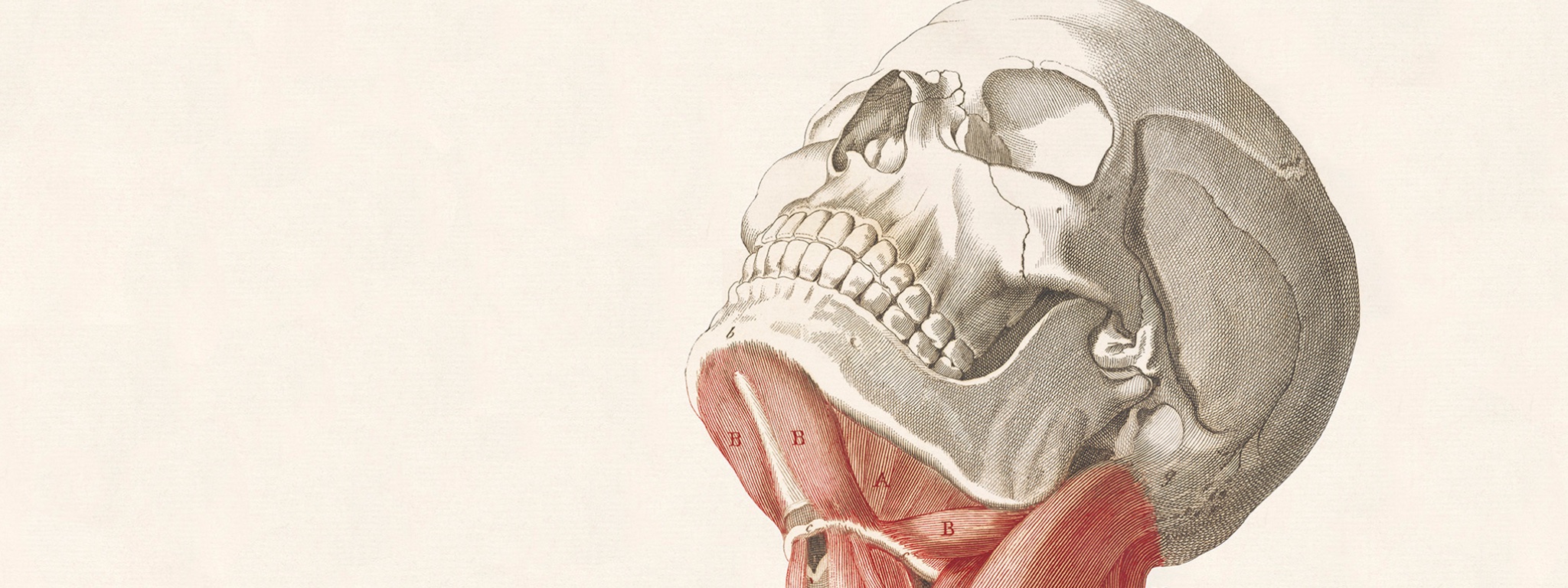
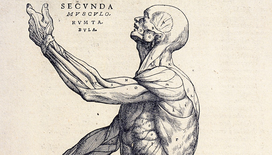
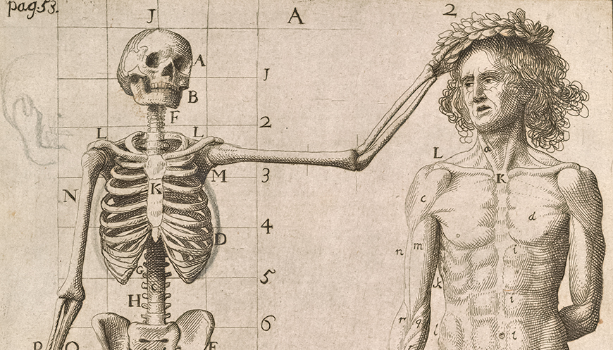
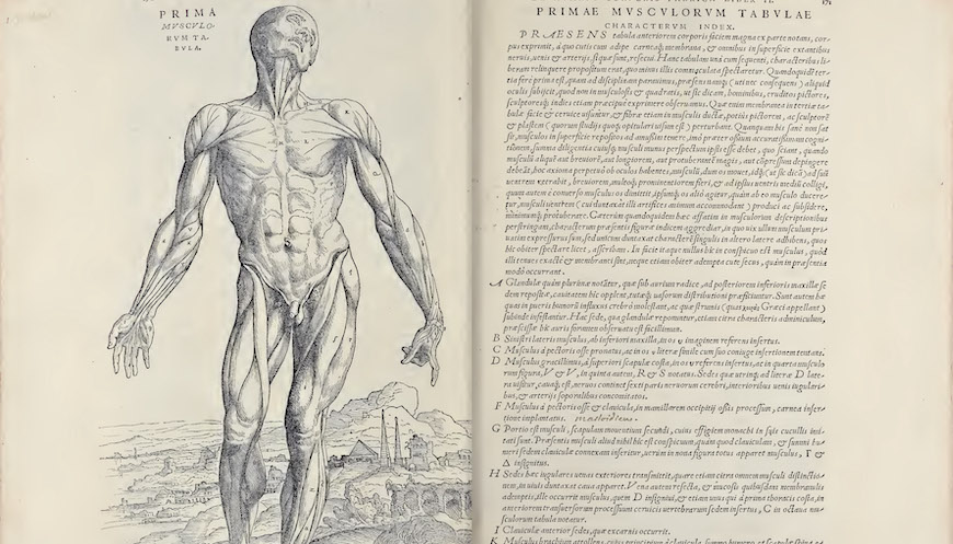
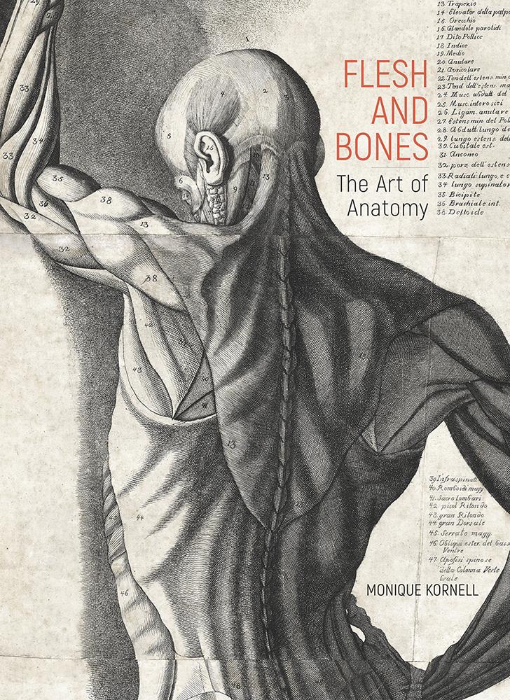
For centuries, the structure of the human body was a fundamental concern for both medicine and art. Anatomy was a basic component of artistic education, and artists were a recognized part of the market for anatomical illustration. At the intersection of art and science, this exhibition looks at the shared vocabulary of anatomical images and at the different methods used to reveal the body through a wide range of media, from woodcut to neon.

This exhibition is presented in English and Spanish. It is one of a series that will test ways to make the presentations in our galleries more welcoming and accessible.

Esta exhibición se presenta en inglés y en español. Es parte de una serie de exhibiciones bilingües que pondrá a prueba varias formas de hacer el contenido que presentamos en nuestras galerías más acogedor y accesible.

SELECTED WORKS

A man stands with his legs apart, holding up large flaps of his dissected skin to reveal his abdominal muscles.

Figure displaying the muscles of the abdomen. From Jacopo Berengario da Carpi, Isagogae breves perlucidae ac uberrimae in anatomiam humani corporis …, 2nd ed. (Bologna, 1523), fol. 7r. Getty Research Institute, 84-B28214

A woodcut illustration, presenting the anatomy of a living body in an antique torso of stone, depicts a dissected abdomen. The intestines have been moved to the side and there is a detailed view of the gall bladder.

Abdominal dissection with detail of the gall bladder, after Jan Steven van Calcar. From Andreas Vesalius, De humani corporis fabrica libri septem (Basel, 1543), p. 365 [465]. Getty Research Institute, 84-B27611

An etched scene depicts the instruction of young art students in a Roman academy. Some draw from a skeleton, while others eagerly observe a dissection in progress in the background.

Academy of Painters, about 1600, Pietro Francesco Alberti. Getty Research Institute, 2721-731

An engraved illustration depicts a partially dissected head that displays the muscles of the eye and the path of the optic nerves to the brain. The left eye, suspended by its muscles, droops downward.

The Muscles of the Eye in its Natural Position and the Muscle of the Eyelid, Shown Separately. From Thomas Bartholin, Anatome ex omnium veterum recentiorumque observationibus, 5th rev. ed. (Leiden, 1686), p. 507, pl. 9. Getty Research Institute, 93-B10050

Two disembodied legs, whose top halves have been dissected down to the knees, walk towards the viewer. A distant town and sailing ships are visible in the distance.

Dissected legs walking in a landscape, by 1616, Francesco Valesio after Odoardo Fialetti. From Giulio Casseri, Tabulae anatomicae, book 4, pp. 90-91, pl. 31, in Adriaan van den Spiegel, Opera quae extant, omnia (Amsterdam, 1645). Getty Research Institute, 84-B2833

In an engraved tableau, five fetal skeletons are arranged on a pile of various stones removed from the body. One appears as a musician playing an instrument made of tissue and bone.

Tableau of fetal skeletons, Cornelis Huyberts. From Frederik Ruysch, Thesaurus anatomicus tertius (Amsterdam, 1703), pl. 1. Getty Research Institute, 94-B12267

A human figure with its skin removed to reveal its muscles stands on a pedestal with its left hand raised above its head. Each body part is labeled with numbers corresponding to text on the left.

Life-size anatomical figure, seen from the front (one of a set of three) (detail), 1780, Antonio Cattani. Getty Research Institute, 2014.PR.17

A human figure with its skin removed to reveal its muscles stands on a pedestal with its left hand raised above its head. Each body part is labeled with numbers corresponding to text on the left.

Life-size anatomical figure, seen from the front (one of a set of three), 1780, Antonio Cattani. Getty Research Institute, 2014.PR.17

A standing woman has been dissected from the chest down, revealing colorful muscles, veins, arteries, and abdominal organs.

Life-size dissected woman (detail), 1750, Jacques Fabien Gautier Dagoty. From Jacques Fabien Gautier Dagoty, Anatomie générale des viscères en situation, de grandeur et couleur naturelle, avec l'angeologie, et la neurologie de chaque partie du corps humain (Paris, [1752]), pls. 1–3. The Huntington Library, Art Collections, and Botanical Gardens, San Marino

A standing woman has been dissected from the chest down, revealing colorful muscles, veins, arteries, and abdominal organs.

Life-size dissected woman, 1750, Jacques Fabien Gautier Dagoty. From Jacques Fabien Gautier Dagoty, Anatomie générale des viscères en situation, de grandeur et couleur naturelle, avec l'angeologie, et la neurologie de chaque partie du corps humain (Paris, [1752]), pls. 1–3. The Huntington Library, Art Collections, and Botanical Gardens, San Marino

In an engraved illustration printed in black and red ink, the head of a classical statue of Apollo appears in a frontal and side view. On the left frontal view, muscles in red are superimposed on the head, while on the right side view, a skull can be seen inside the outline of the head, which retains its distinctive topknot hairstyle.

The head of the Apollo Belvedere, rendered anatomically, Nikolaj Utkin after Jean Galbert Salvage. From Jean Galbert Salvage, Anatomie du gladiateur combattant, applicable aux Beaux Arts (Paris, 1812), pl. 1. Getty Research Institute, 85-B12146

A lithographic illustrated plate depicts the upper half of a male body, head turned to the right, eyes closed, his dissected chest and abdomen on display, the vascular system picked out in light pink and blue letters and fragments of words in different colors mounted upright on a wood base.

Dissection of chest and abdomen, Thomas Sinclair after Joseph Maclise. From Joseph Maclise, Surgical Anatomy (Philadelphia, 1851), pl. 26. Getty Research Institute, 1373-163

Two images of a human figure, one facing forward and one facing back, stand side by side in a foldable box. The forward-facing figure on the left has 33 detachable anatomical parts made of color-printed steel.

Smith's New Outline Map of the Human System, 1888, American Manikin Co. Peoria, IL. Biomedical Library. Library Special Collections for Medicine and the Sciences, Louise M. Darling Biomedical Library, UCLA

A male nude stands with arms raised and head cropped out, his body slightly facing left with the left leg forward and his torso twisting to the front. Shadows from window blinds are cast along his body.

Ken Moody, negative 1985, print 2011, Robert Mapplethorpe. Gift of the Robert Mapplethorpe Foundation to the J. Paul Getty Trust and the Los Angeles County Museum of Art. © Robert Mapplethorpe Foundation. 2012.52.10

Pulsating neon tubes illuminate a human body's circulatory system as it floats against a black backdrop.

Robert, 2018, Tavares Strachan. Photo: Andrea D’altoè Neonlauro. Photo courtesy the artist and Marian Goodman Gallery, New York

Figure displaying the muscles of the abdomen. From Jacopo Berengario da Carpi, Isagogae breves perlucidae ac uberrimae in anatomiam humani corporis …, 2nd ed. (Bologna, 1523), fol. 7r. Getty Research Institute, 84-B28214

RELATED EVENTS

Under the Skin: Drawing Anatomy

March 12, 2022
Getty Center

The Polykleitos Problem: Illusions of the Ideal in European Anatomical Images

June 24, 2022
Getty Center and Online

RELATED PUBLICATION

Flesh and Bones: The Art of Anatomy

Monique Kornell

GALLERY TOURS

Thursdays at 2:00 p.m.
March 10–July 7, 2022
(Note: no tours on March 17, 31, and April 7, 28)

Space is limited. Please sign up at the Research Institute lobby desk

MOBILE TOUR

Explore this exhibition with a free audio tour, available in English and Spanish. Download the GettyGuide® app

EXHIBITION DOCUMENTATION

Get Tickets