7. Evaluating Multiband Reflectance Image Subtraction for the Characterization of Indigo in Romano-Egyptian Funerary Portraits
The Brooklyn Museum (BKM) began acquiring Egyptian antiquities in 1902 and now boasts one of the largest holdings of Egyptian materials in the United States. This renowned collection contains six Romano-Egyptian funerary portraits on : Portrait of Demetrios (11.600, AD 95–100), Woman with Earrings (1996.146.9, AD 100–105), Mummy Portrait of a Man (ca. AD 120–130), Noblewoman (ca. AD 150), Boy with a Floral Garland in His Hair (ca. AD 200–230), and Portrait of a Young Person (ca. AD 200–230). Two of these portraits are characterized as paintings with aqueous and four as paintings with wax binding media. Aside from the Noblewoman portrait (fig. 7.1), which has been extensively restored, the portraits survive in remarkably good condition. All six portraits were documented and analyzed through visual examination, , , , , , , and .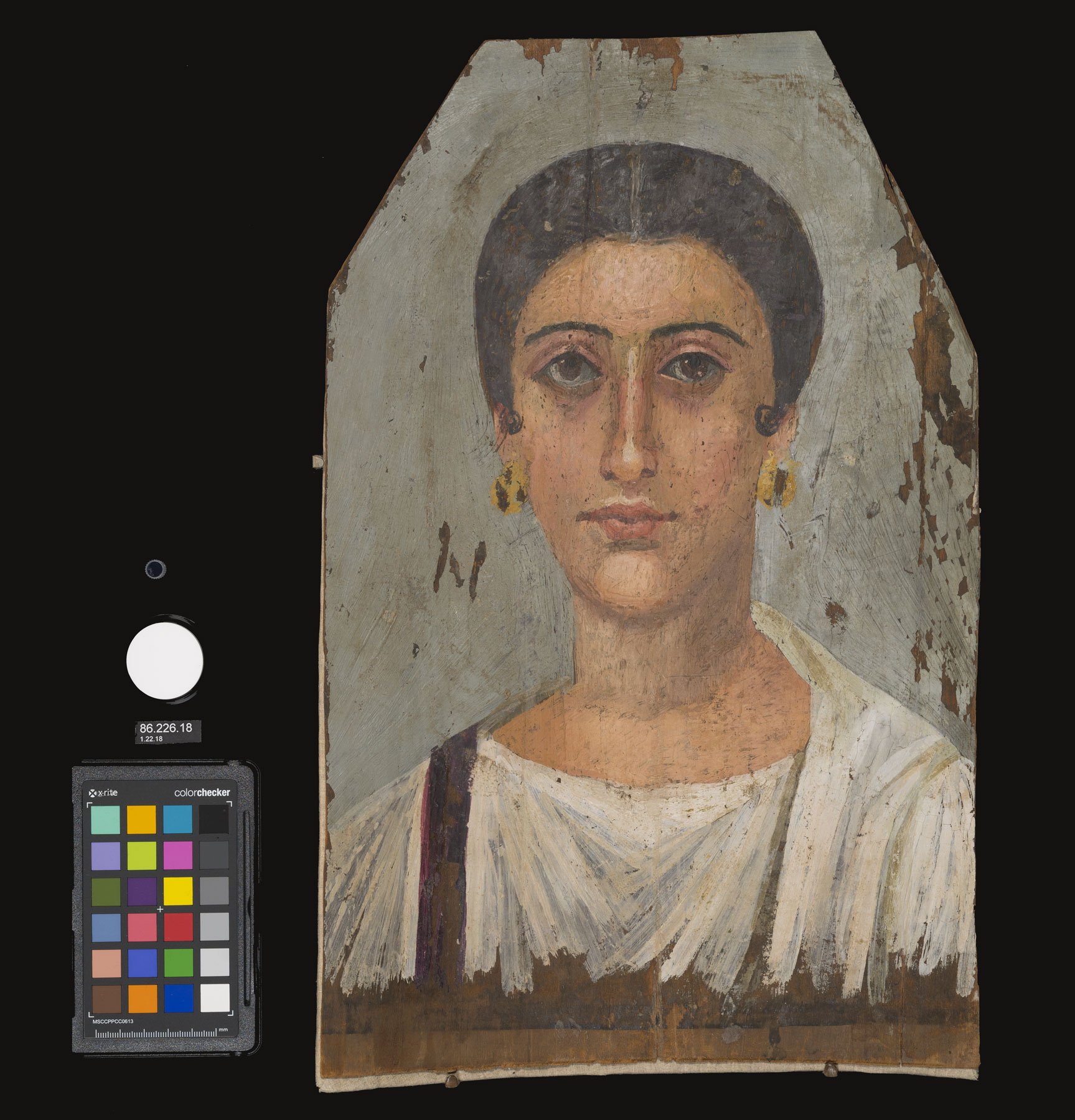 Figure 7.1
This paper focuses on for the characterization of . As few references to this technique exist in the literature, BKM conservators not only evaluated the information gained from its application to the study of Romano-Egyptian funerary portraits but also investigated the technique itself, refining variables in image capture and processing to optimize results. Protocols were developed for equipment setup and image capture using reflectance standards, color standards, and material samples as internal references. Spectral curves collected using FORS elucidated why materials other than indigo may be visualized in processed subtraction images. Selected data obtained from MBI, FORS, and Raman spectroscopy are discussed in this paper, along with relevant XRF results.
Indigo was detected exclusively in mixtures with a red on three of the encaustic portraits; indigo, both in mixtures and alone, was found more widely on the two tempera portraits. Indigo was not found on the sixth portrait, also encaustic.
The use of indigo or woad on Egyptian textiles dates back to as early as the sixteenth century BC.1 Woad (Isatis tinctoria) was more commonly used as a dye; indigo (Indigofera tinctoria), likely imported from India starting in the Ptolemaic era,2 was more commonly employed as a pigment.3 The term indigo is used throughout this paper to encompass indigotin-based colorants irrespective of plant source.
Pioneered by Webb and colleagues,4 the MBR technique described in this essay combines one near-infrared image and one visible light image in digital post-processing. This noninvasive and nondestructive technique can visualize and localize materials, including indigo, producing a surface map.
To evaluate our modifications to and application of the imaging technique, analyses were carried out using a visible–near infrared fiber optics reflectance spectrometer and a handheld Raman spectrometer. In addition, samples were taken for analysis using a benchtop Raman instrument.
Paint-out boards created using historically consistent materials were imaged and analyzed with FORS, serving as simplified analogues of the portraits and providing references for known materials in mixtures with indigo.
Multiband Imaging (MBI)
MBI image suites—including visible, , , IRR, and images—of each portrait and paint-out board were captured using a modified Nikon D610 DSLR camera with the UV/IR filters removed and a Jenoptik 60 millimeter UV-VIS-IR APO Macro lens (see fig. 7.2 for lighting and filter specifications). and images were generated by combining reflectance captures via channel substitution in Adobe Photoshop.5 A Spectralon 99 percent reflectance standard from Labsphere,6 an X-Rite ColorChecker Passport, and an unbound indigo pigment sample7 were included in all captures (see fig. 7.1). An unbound pigment sample was included in all VIL captures. MBI suites of the paint-out boards included dry samples of the binding media and pigments used on each board (fig. 7.3).
| Light Source | MBI Type | Filters |
|---|---|---|
Genaray Spectro LED-14 Lights (Output: 5600K) | Visible (VIS) | IDAS-UIBAR filter (375–700 nm bandpass) |
UV Systems LW370 TripleBright II Lights (Output: 368 nm, 5750K) | Ultraviolet-induced visible fluorescence (UVF) | IDAS-UIBAR filter Kodak 2E pale yellow optical Wratten filter (410 nm longpass) |
Ultraviolet reflectance (UVR) | X-Nite BP1 filter (320–670 nm bandpass) X-Nite 330 filter (270–375 nm bandpass) | |
Solux Halogen MR-16 Lights (Output: 4700K) | Infrared reflectance (IRR) | X-Nite 830 filter (830 nm longpass) |
Captures taken to be processed for multiband reflectance image subtraction (MBR) | MidOpt BP660 filter (640–680 nm bandpass) MidOpt BP735 filter (715–780 nm bandpass) | |
American DJ RGB LED Lights, Model 64B LED PRO, Red Bulbs CL1 (Output: 629 nm) | Visible-induced infrared luminescence (VIL) | X-Nite 830 filter (830 nm longpass) |
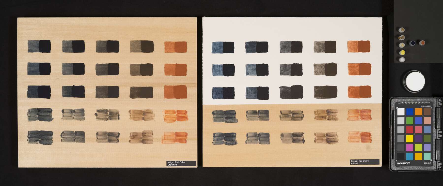 Figure 7.3
Multiband Reflectance Image Subtraction (MBR)
MBR to localize indigo was performed by illuminating the field with Solux MR-16 halogen bulbs, 4700 Kelvin output, and taking two captures: one with a MidOpt BP660 bandpass filter and one with a MidOpt BP735 bandpass filter. Captures were converted to grayscale in Adobe Photoshop’s Camera Raw utility by setting the saturation to -100, and saved as TIFFs.8 The desaturated TIFF images were then combined using the “Difference” Blend Mode in Photoshop to generate a subtraction image. Processing could alternatively be carried out using free and open-source software such as ImageJ and GIMP.
The “Difference” function takes the absolute value of the difference between two source images, pixel by pixel, to generate a new image. Corresponding source-image pixels with similar RGB values yield small numerical differences, while the combination of pixels with dissimilar RGB values creates large numerical differences. Differences of zero in each color channel result in a black pixel, while differences of increasing magnitude generate pixels approaching white as the values approach 255 in each channel. Materials with little change in reflectance across the spectral regions encompassed by the BP660 and BP735 filters result in small numerical differences and appear dark in the MBR image.
Characteristic reflectance spectra for indigo display strong absorbance around 660 nanometers and strong reflectance just under 800 nanometers. The pairing of narrow bandpass filters centered at 660 nanometers and 735 nanometers exploits the pronounced difference in indigo’s reflectance in the visible and near infrared (fig. 7.4).9 Indigo in various forms, including Maya blue, yields large numerical differences and appears bright in MBR images generated from captures made using these filters. Imaging performed at BKM indicates that other blue materials including lapis lazuli and ultramarine; cobalt-containing blues, such as cobalt blue, smalt, and cerulean blue; and, to a lesser extent, Egyptian blue, can also produce brightness in these images due to their reflectance behaviors.10 MBR as a characterization technique is strengthened in combination with knowledge of an object’s historical context and through corroboration by other imaging and analytical methods.
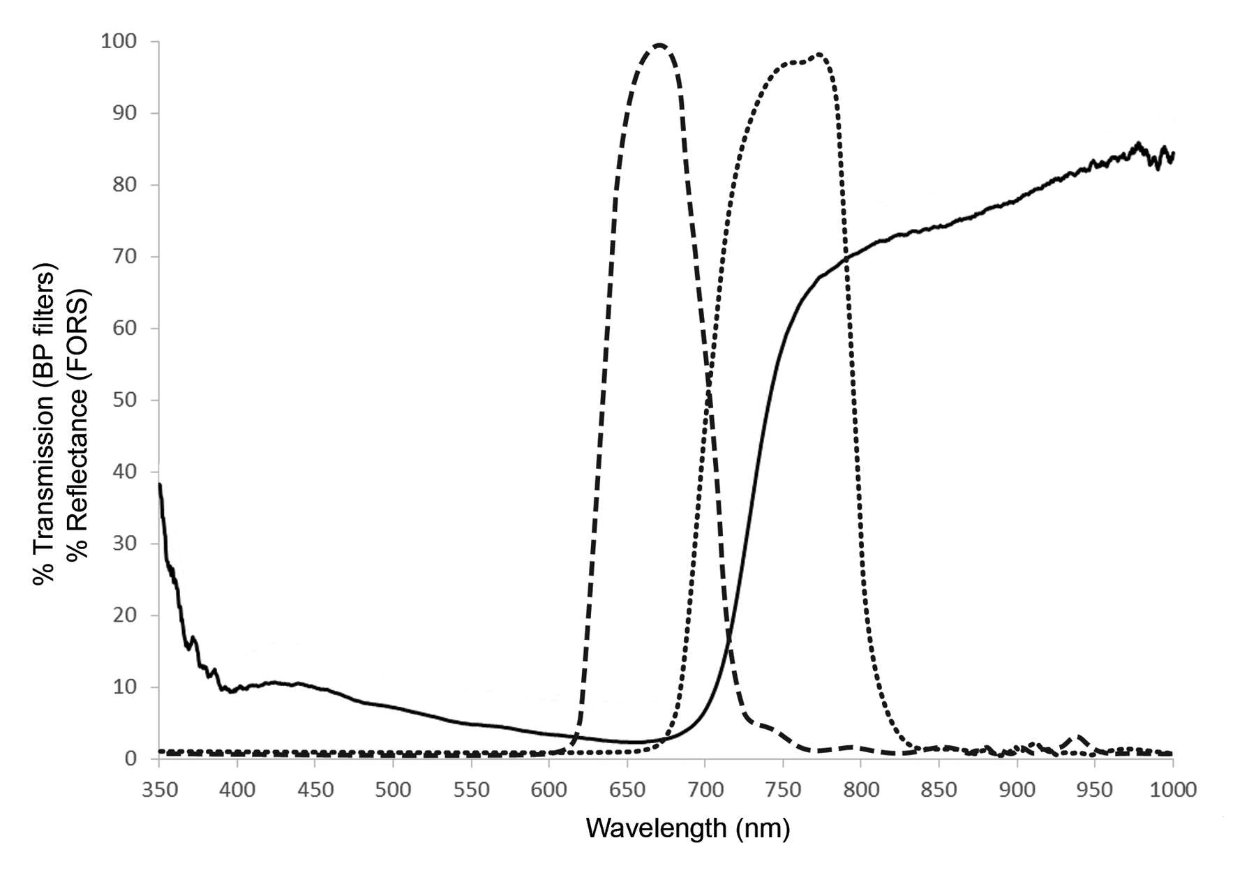 Figure 7.4
To achieve reliable and consistent MBR results, the measured exposure of the BP660 capture should be as close as possible to that of the BP735 capture without exceeding it.11 Because the two bandpass filters pass different amounts of light, the BP660 capture typically requires a longer exposure time than that of the BP735 capture to result in a pair of images in which the BP660 capture is as close as possible to but still darker than the BP735 capture. Ensuring that the exposure gap between the two captures is as narrow as possible maximizes the specificity of the MBR technique, highlighting those materials with the largest differences in reflectance in this spectral region. The exposure of each capture was assessed using the RGB values of the Spectralon reflectance standard and the Neutral 8 gray square on the X-Rite ColorChecker Passport. The ColorChecker gray square was used because the American Institute for Conservation imaging guidelines already utilize this standard, recommending an RGB value of 200 in both visible and infrared photography.12
As with many imaging techniques, shifts in camera position or lighting between captures and uneven lighting can confound processing and undermine the usefulness of reflectance and color standards. Unlike with many other imaging techniques, suboptimal captures not only lower the quality of MBR results but can actually create misleading or erroneous images. Capture sets where one or both relative exposure values were higher in the BP660 shot were empirically found to produce erroneous results, in which some materials appeared bright or dark in ways not clearly related to each other or to known reflectance behaviors. Capture sets with the desired arrangement of exposures but larger exposure gaps produced MBR images with wider grayscale ranges, reducing the specificity of the technique. Adjusting exposures post-capture utilizes nonlinear functions and can also yield unrepresentative results.
Raman
Raman analysis of samples taken from the portraits was performed microdestructively using a benchtop Bruker Senterra Raman spectrometer equipped with a 50x microscope objective and a charge-coupled device (CCD) detector. A continuous-wave diode laser emitting at 785 nanometers was used as the excitation source, and two holographic gratings (1800 and 1200 rulings per mm) provided a spectral resolution of 3 to 5 reciprocal centimeters. The output laser power, number of scans, and integration time were adjusted based on the Raman response of the sample being analyzed.
In situ Raman analysis was conducted nondestructively using a handheld Bruker Bravo Raman spectrometer equipped with a CCD detector, featuring double laser excitation (785 nm and 852 nm) and providing a resolution of 10 to 12 reciprocal centimeters. The output laser power was approximately 50 milliwatts for both lasers, while the number of scans and integration time were adjusted based on the Raman response of the area being analyzed. Spectra were interpreted by comparison with the Metropolitan Museum of Art’s library databases and with published literature.
Fiber Optics Reflectance Spectroscopy (FORS)
FORS readings were taken using an Ocean Optics FLAME-S-UV-VIS-ES spectrometer with an instrument range of 350 to 1000 nanometers, a cable range of 400 to 2100 nanometers, and a full-width, half-maximum optical resolution of approximately 1.5 nanometers. Spectra were recorded using OceanView software, and data were interpreted and plotted using Microsoft Excel. Three to five readings were taken for each color analyzed.
X-Ray Fluorescence Spectroscopy (XRF)
XRF readings were taken using a handheld Bruker Tracer III-V+ instrument with a rhodium source, a beryllium sample window roughly 3 by 4 millimeters, and a SiPIN detector. Two readings were taken at each spot: one at 40 kiloelectron volts, 3 microamps, 60 seconds, and one at 15 kiloelectron volts, 32 microamps, 60 seconds under vacuum to improve sensitivity to lower-mass elements. Spectral data were acquired and interpreted using S1PXRF and Artax software.
Paint-Out Boards
A set of reference paint-out boards was made as a guide to better understand how indigo-containing paint films respond to MBR (see fig. 7.3). The materials used to make the boards were selected based on a literature review of Romano-Egyptian painting practices, on material characterizations in the APPEAR database, and on FORS and XRF analyses of the BKM portraits. Five binding media and seven pigments in addition to indigo, all unaged, were chosen.13
Linden (Tilia americana) panels were used as the support wood. In the APPEAR database, portraits described as having an aqueous binder usually have a white layer, while those described as wax commonly have no ground, or sometimes a black ground. The white ground on many aqueous portraits, including figure 7.5, is thickly applied with a visible brush texture.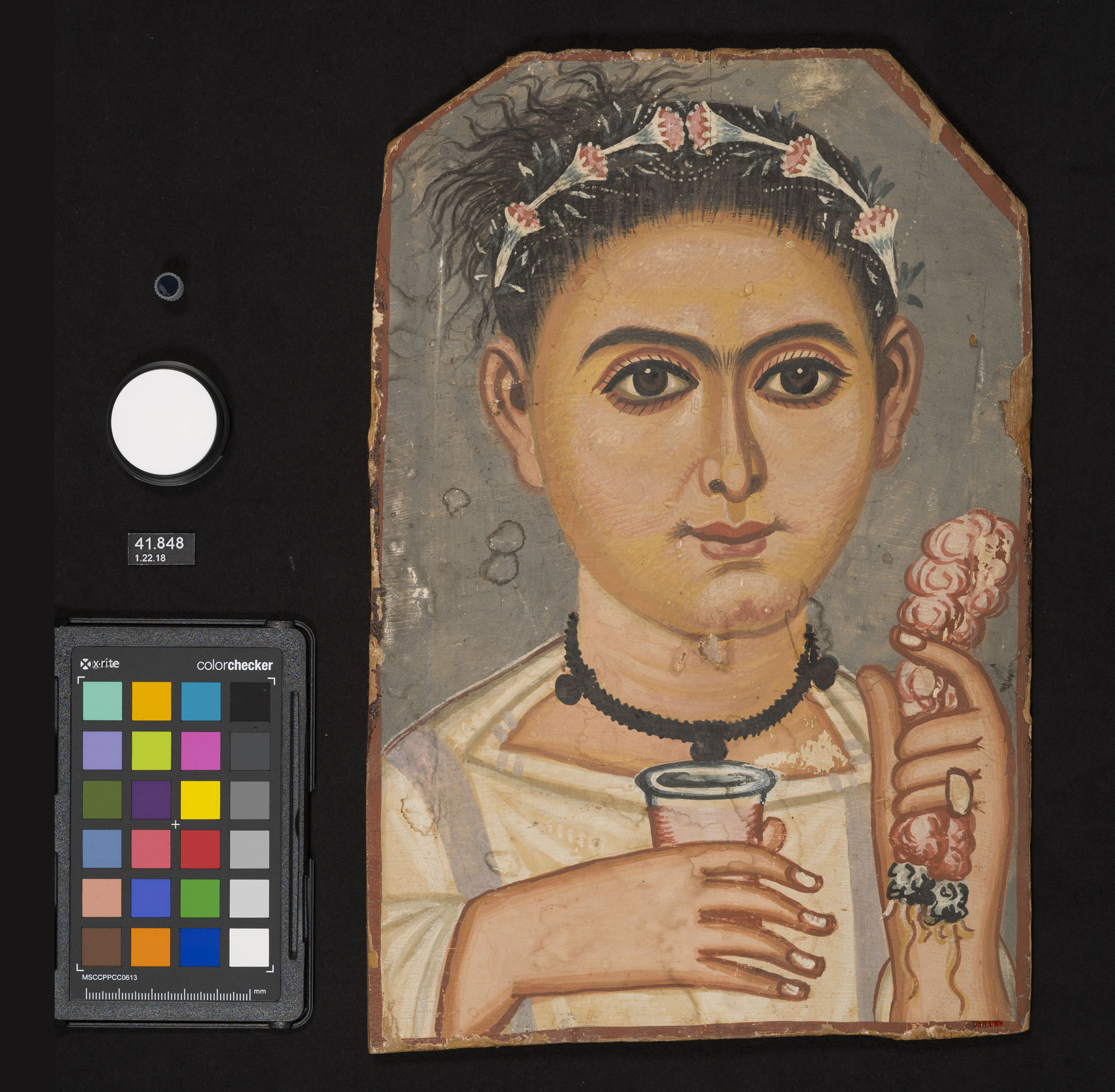 Figure 7.5
One set of panels was sized with two layers of cowhide glue followed by a partial ground layer of bound in cowhide glue, applied to mimic the brush texture observed on the portraits. The other set of panels was left unprepared as a comparative control.
On each board, gradients between indigo and one other pigment were established using three aqueous binders—one cowhide glue and two rabbit-skin glues—and two binders—one yellow and one white. The pigments include Egyptian blue, , , , gypsum, , and vine black. On the prepared boards, the aqueous paint-outs were applied over the gypsum ground and the wax paint-outs were applied directly on the sized wood. Each gradient step was divided into two halves. For the aqueous paint-outs, the halves comprise a single and a double layer of the same paint, while for the encaustic paint-outs, each half represents the same amount of wax combined with more or less pigment.
Indigo has a high tinting strength and was found on many of the BKM portraits in mixed pale purple hues that would have required only small amounts of colorant to create. To better represent the hues observed in the portraits, an additional pair of boards was made using minute quantities of indigo mixed with red ochre or madder.
Results
Indigo was identified on five of the six portraits in the BKM collection using FORS and Raman in combination with MBR images. On the three encaustic paintings in which it was identified, indigo was found exclusively in mixtures with a red lake pigment. On the two tempera portraits, indigo was detected more extensively throughout and in a broader range of color mixtures.
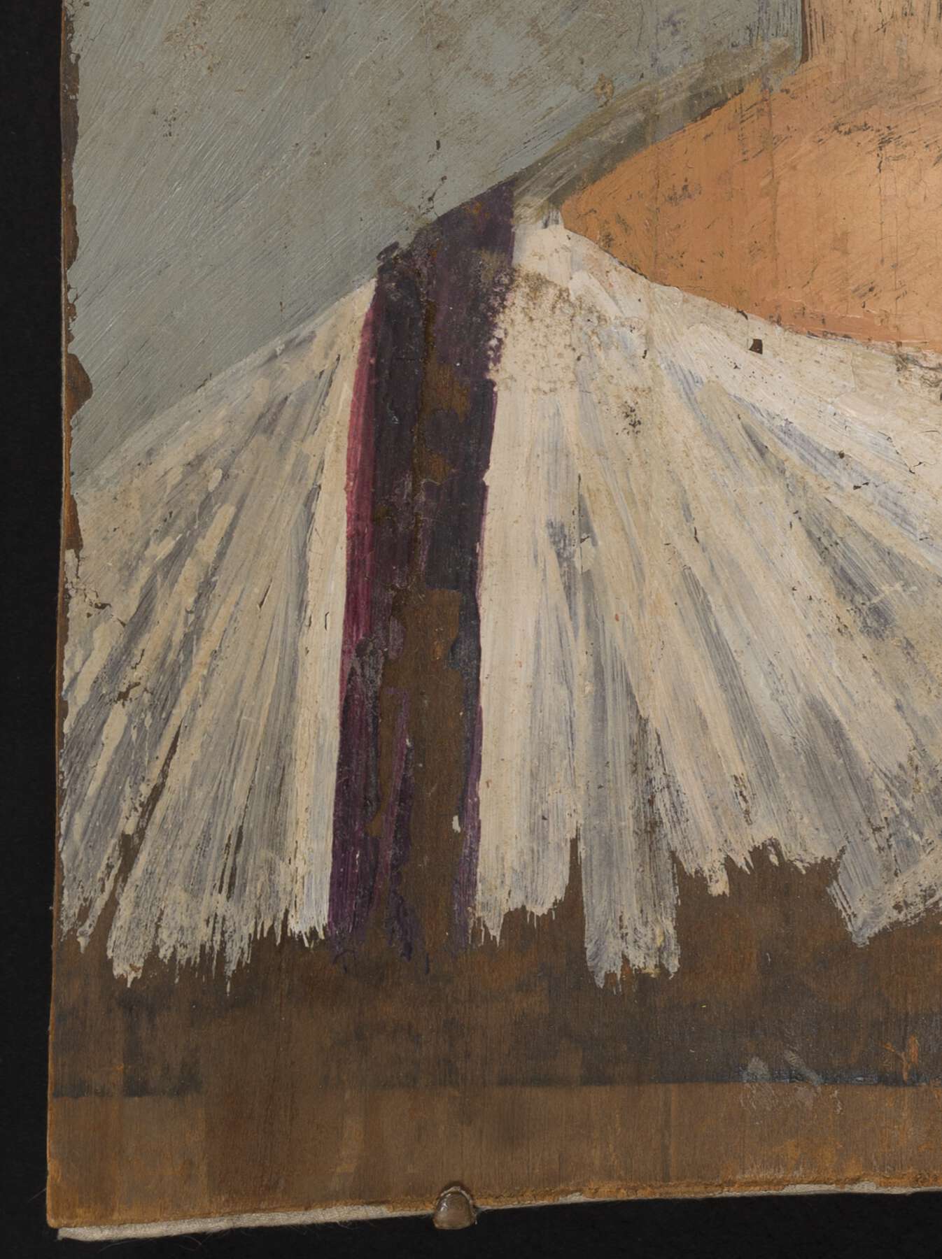
On the encaustic portraits of Demetrios and the Noblewoman (see fig. 7.1), the were rendered using indigo-containing paints applied to different effects. On Demetrios, a rich, dark purple layer was applied over a lighter pink layer. On the Noblewoman, overlapping brushstrokes of purple and pink paints with varying opacities were used. Indigo was identified in the purple paints on both portraits, but not in the pinks (fig. 7.6).14 A red lake pigment, most likely madder,15 comprises the dominant pigment in the pink paint and is mixed with indigo to make the purple.
 a. Normal light
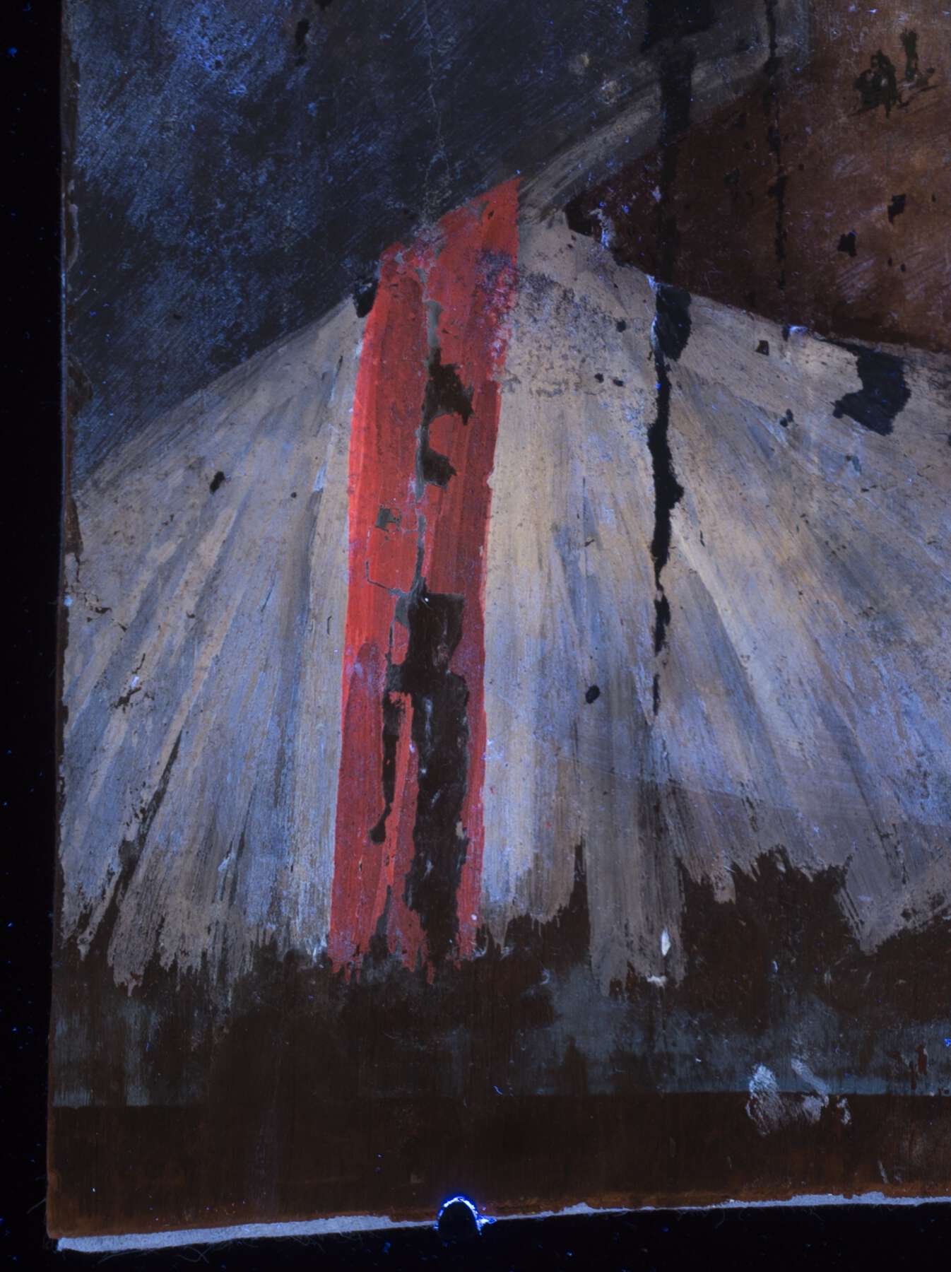 b. UVF
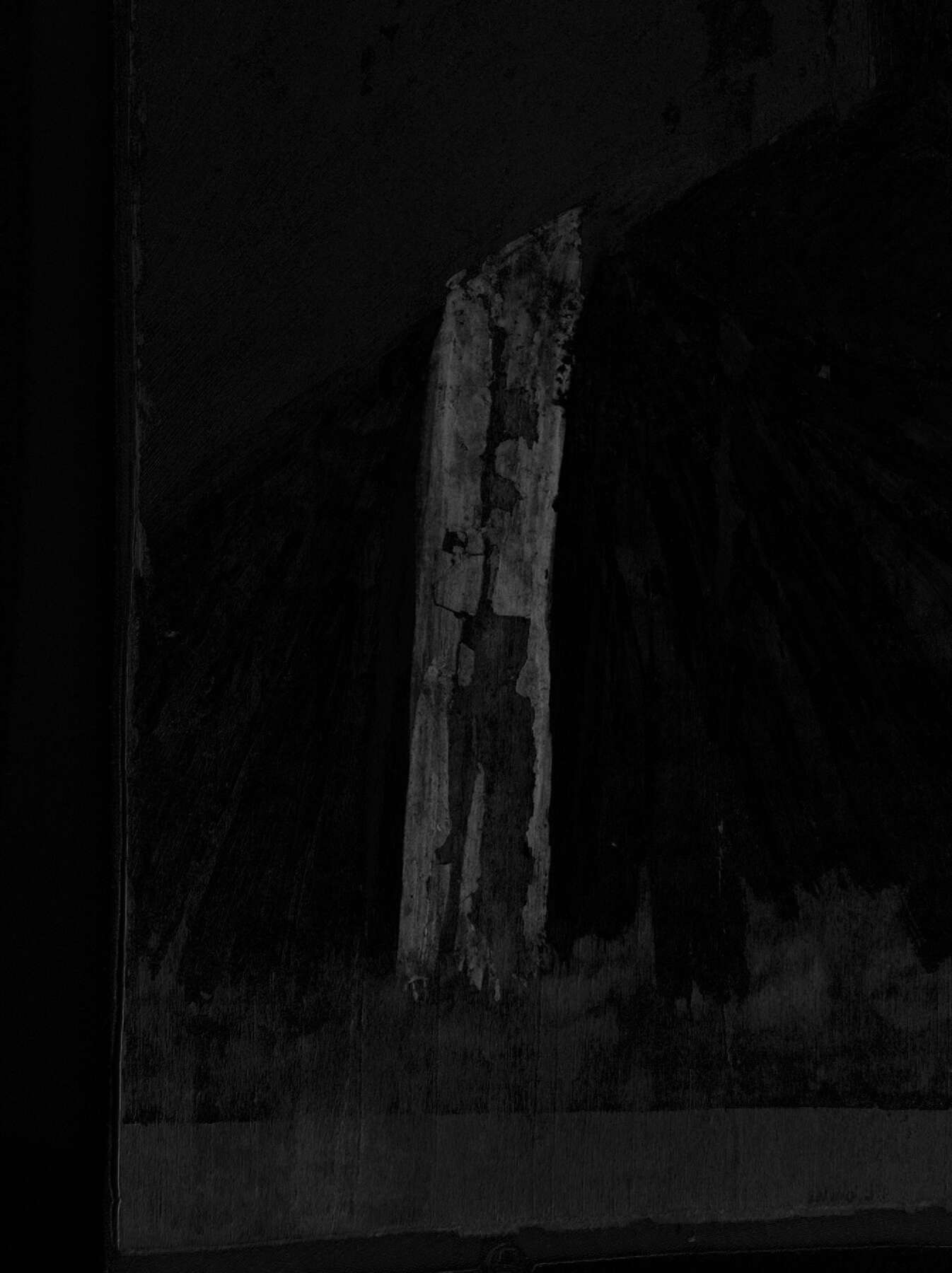
The Mummy Portrait of a Man is an encaustic portrait that retains remnants of funerary wrappings, including resin, textile, and white cartonnage painted with tempera (fig. 7.7). This portrait has two clavi: one rendered in encaustic and painted at the same time as the sitter’s face, and one in tempera that was added on top of the cartonnage when the panel was integrated into the mummy bundle. The encaustic clavus was ultimately covered by wrappings. Indigo was found only in the purple of the tempera clavus and was mixed with a red lake pigment (fig. 7.8). XRF, FORS, and Raman spectroscopy found no evidence of a blue pigment in the blue-gray encaustic clavus.
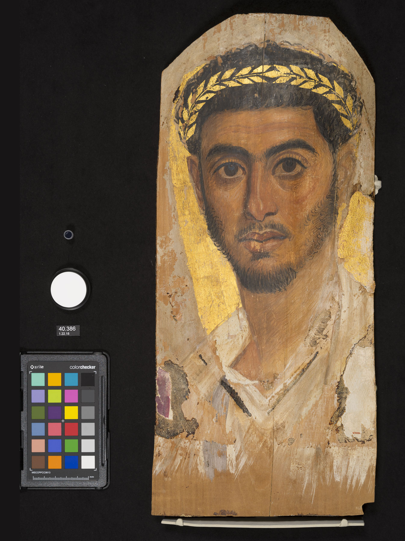 Figure 7.7
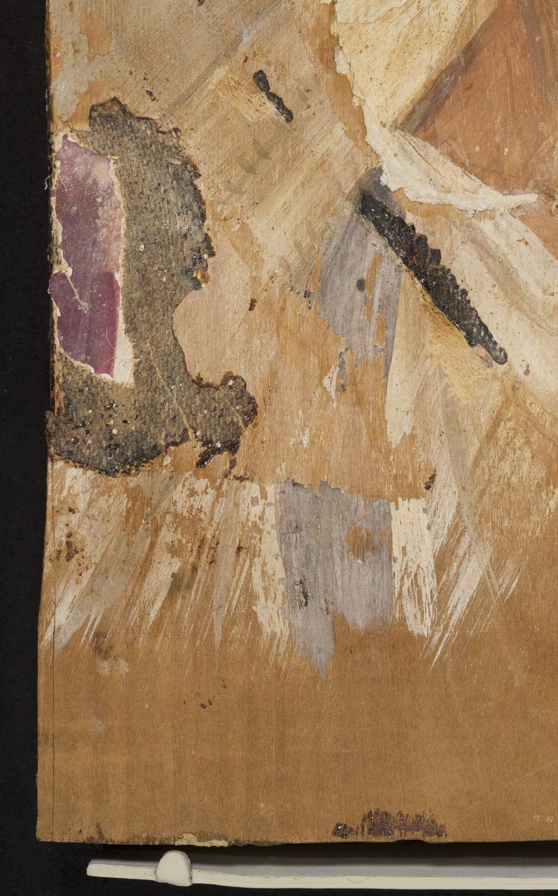 a. Normal light
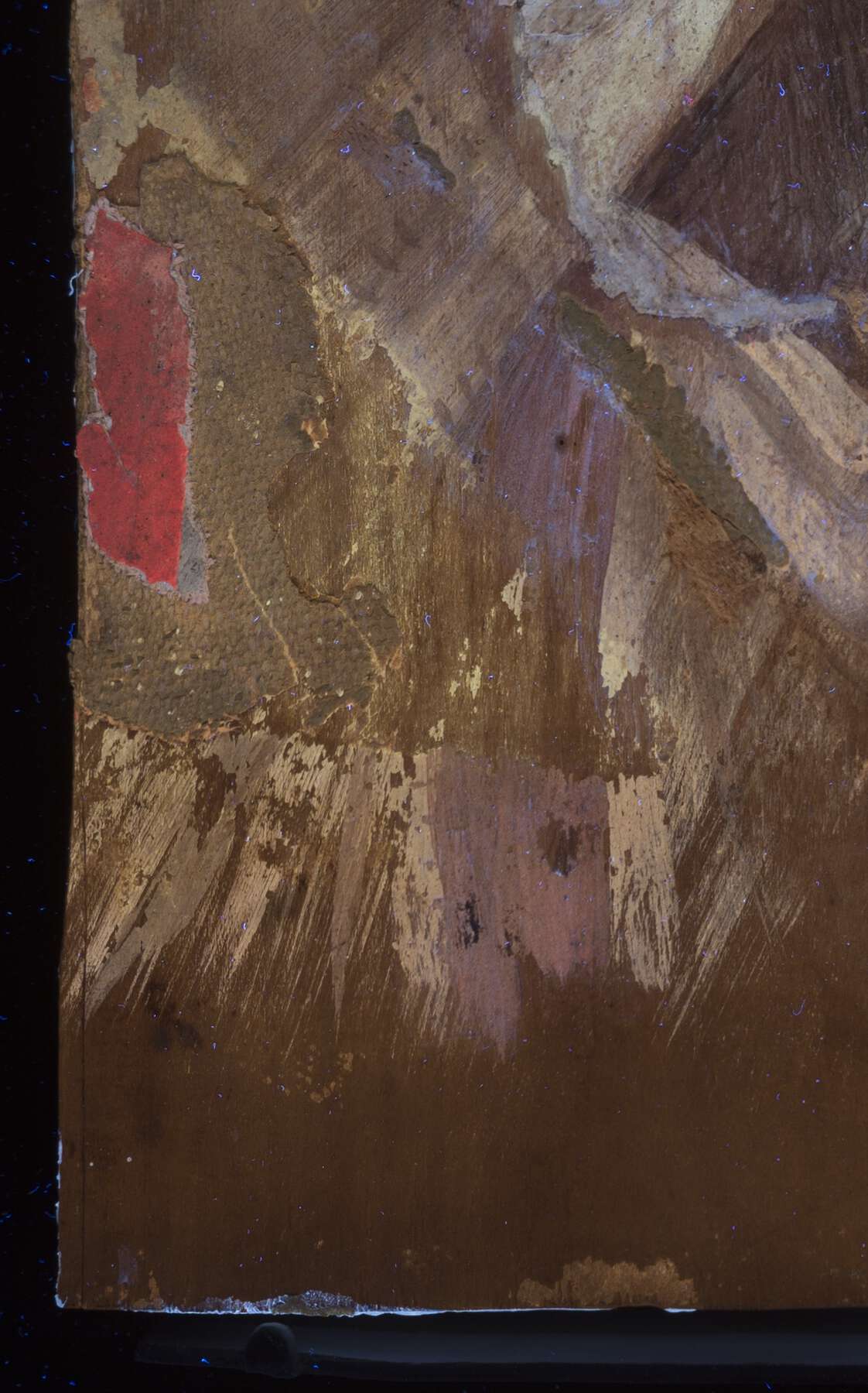 b. UVF
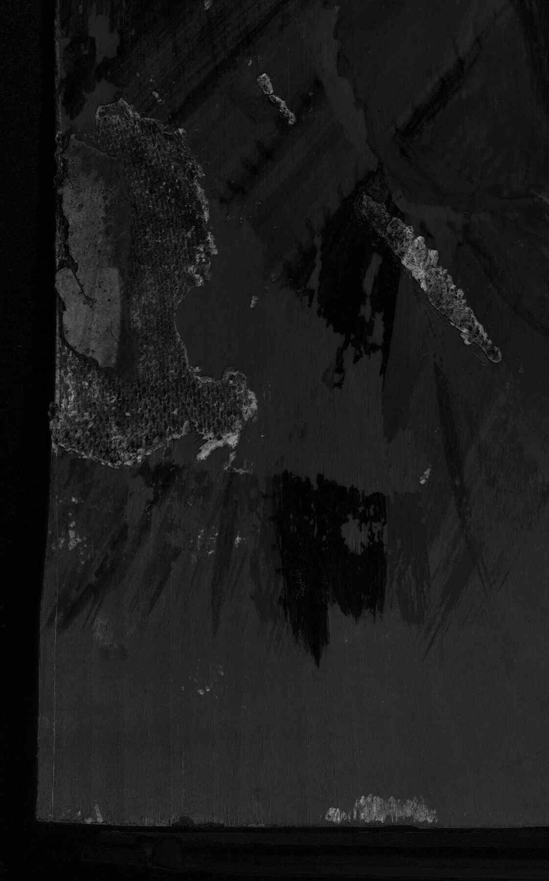
On the tempera portraits, indigo was identified in mixtures, creating both blue and purple colors. Indigo was used as the dominant pigment to paint bluish details including the rims of the cups and the bases of the held by both sitters. On the Portrait of a Young Person (fig. 7.9), indigo was also detected in the dark blue decorative bands on the neckline of the undertunic. On the Boy with a Floral Garland in His Hair (see fig. 7.5), indigo was found in the leaves and flowers of the floral crown, as well as in the decorative bands and the clavus, where it was mixed with a red lake pigment to create a pale purple hue (fig. 7.10). These regions showed a very faint MBR response, appearing light pink in FCIR and light blue in FCUV. These false-color responses are similar to those observed on the paint-outs made as a reference for the pale purple hue using a small quantity of indigo mixed with madder.
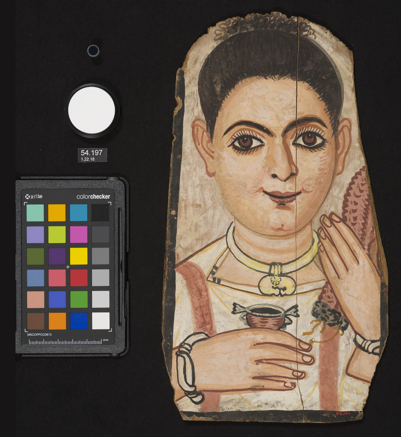 Figure 7.9
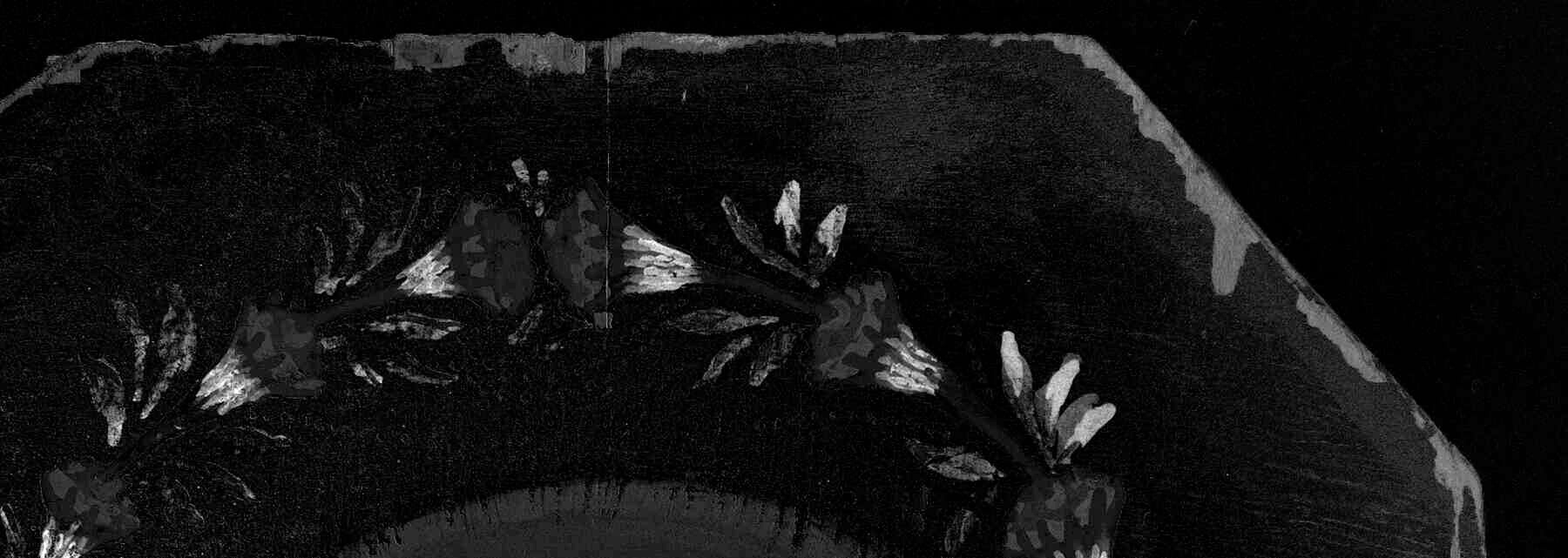 Figure 7.10
Discussion
The use of multiple techniques clarified what data each method can yield individually and what information can be gleaned when the results are considered together. FORS analysis, which essentially exploits the same reflectance phenomena as MBR, provided localized spectral information that corroborated the material responses observed in the MBR images and elucidated the circumstances under which the subtraction technique can yield misleading results. Raman analysis was used as a complementary technique to substantiate or challenge material characterizations made using MBI and FORS. The full MBI suite and XRF data were considered in combination with results of these analyses to further characterize the pigments present on the portraits.
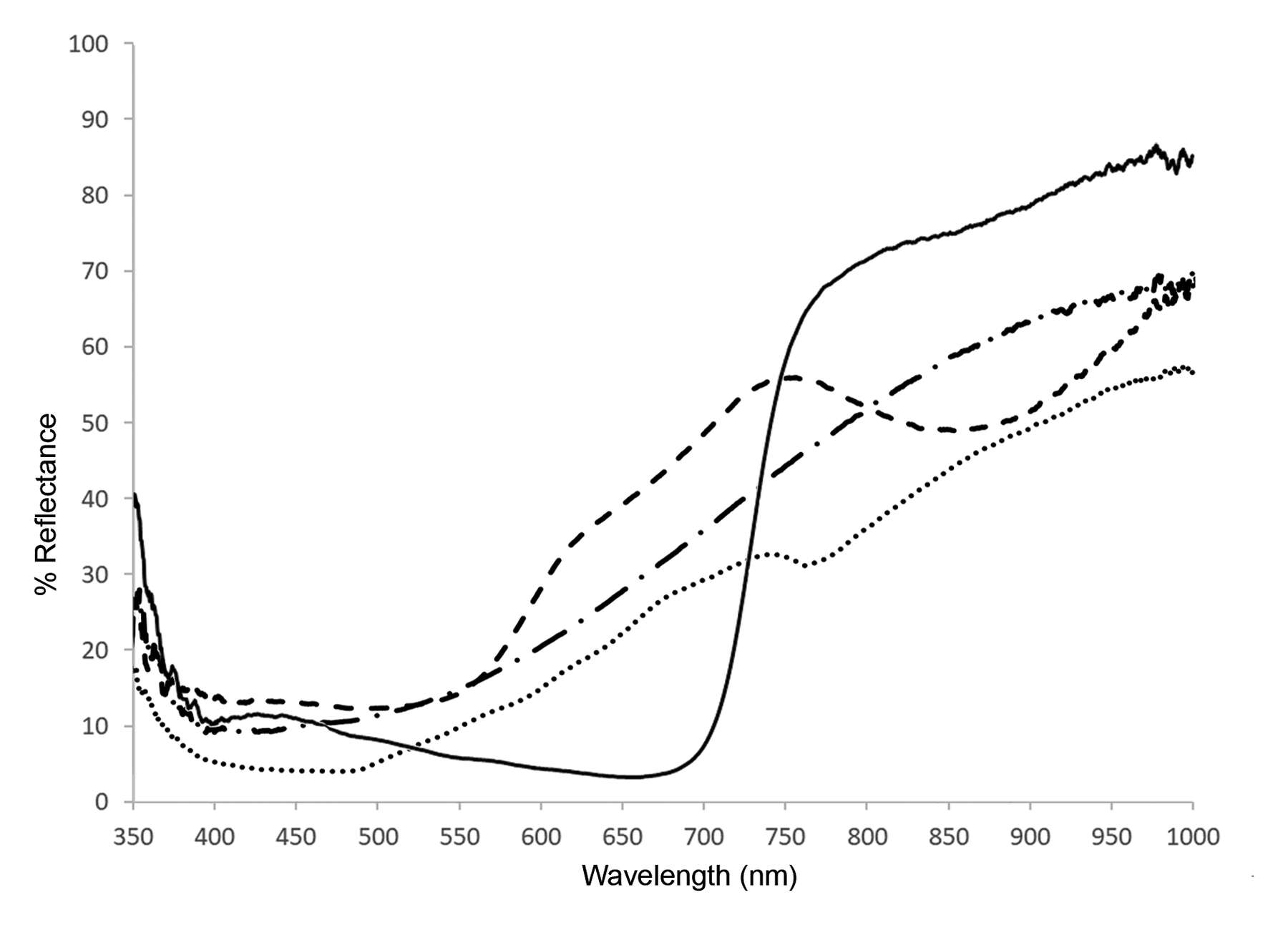 Figure 7.11
Materials that have reflectance behaviors similar to those of indigo, in the range of 660 to 800 nanometers, can result in confounding MBR responses. In Romano-Egyptian mummy portraits, such materials include wood, resin, and red ochre (fig. 7.11). Wood yielded a consistent MBR response, both on unpainted areas of the reference boards and on the portraits where thin paint application or loss exposed the wood support (see fig. 7.10). On the Mummy Portrait of a Man (see fig. 7.7), translucent red-brown resin associated with the mummy wrappings appeared almost as bright as the indigo-containing paint (see fig. 7.8). Red ochre was faintly visualized on the reference paint-outs (fig. 7.12) and on the two tempera portraits in the dark red outlines surrounding the flesh tones and in the dark reds of the garlands (see fig. 7.10). Visual examination combined with MBR imaging could imply the dark purple-red color on the portraits was achieved through an admixture of indigo; however, XRF, Raman spectroscopy, and FORS indicated the color was achieved by red ochre alone.
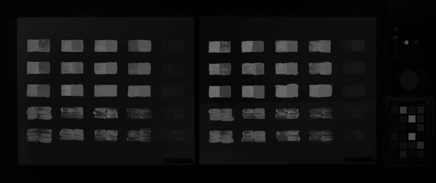 Figure 7.12
Using multiple images from the MBI suite in concert can elucidate the distribution of pigments across a painted surface. In the Boy with a Floral Garland in His Hair (see fig. 7.5), the floral crown was painted using white, blue, pink, and red. The blue leaves and bands on the flowers contain indigo and appear bright in the MBR image. The dark red paint surrounding the center of each flower contains red ochre and appears faintly visible (see fig. 7.10). The pink centers of the flowers appear dark and were painted using a red lake pigment, likely madder. In the absence of other analytical techniques, UVF and FCUV images can help characterize madder and red ochre (figs. 7.13 and 7.14). On the crown, the red lake pigment fluoresces brightly under ultraviolet radiation and appears blue in FCUV images, while the red ochre absorbs, appearing dark in UVF and dark purple in FCUV. These color responses and relationships proved consistent throughout this project, enabling comparisons across portraits, paint-out boards, and reference standards.
 Figure 7.13
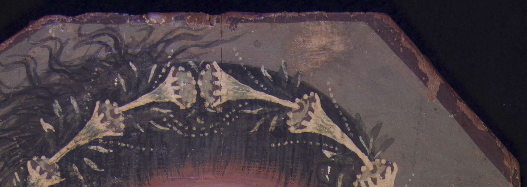 Figure 7.14
Synthesizing the results of multiple techniques provides a richer understanding of material usage and distribution. On the Noblewoman (see fig. 7.1), the hair was rendered by applying an unmodulated layer of paint over the black ground; the hairstyle was then defined by adding a central part and tightly coiled curls at either ear in addition to short, directional highlights. FORS and handheld Raman identified indigo in the curls and highlights; however, MBR yielded no visible response. Images of the reference boards suggest that minute amounts of indigo are difficult to visualize via MBR. The painted details in the hair appear somewhat degraded under magnification; aging of the pigments and/or binding media may be affecting indigo’s MBR response. The hairstyle details fluoresce strongly under UV, consistent with madder (fig. 7.15). Considering the FORS and Raman data together with the UVF image strongly suggests the brushstrokes comprise an indigo-madder mixture. Additionally, the blue-green color of these brushstrokes in FCUV (fig. 7.16) is similar to the FCUV color of the unbound indigo standard included in all images and of indigo mixed with madder on the paint-out boards. Although FCUV alone is not diagnostic for indigo, within this suite of images it is notably corroborative.
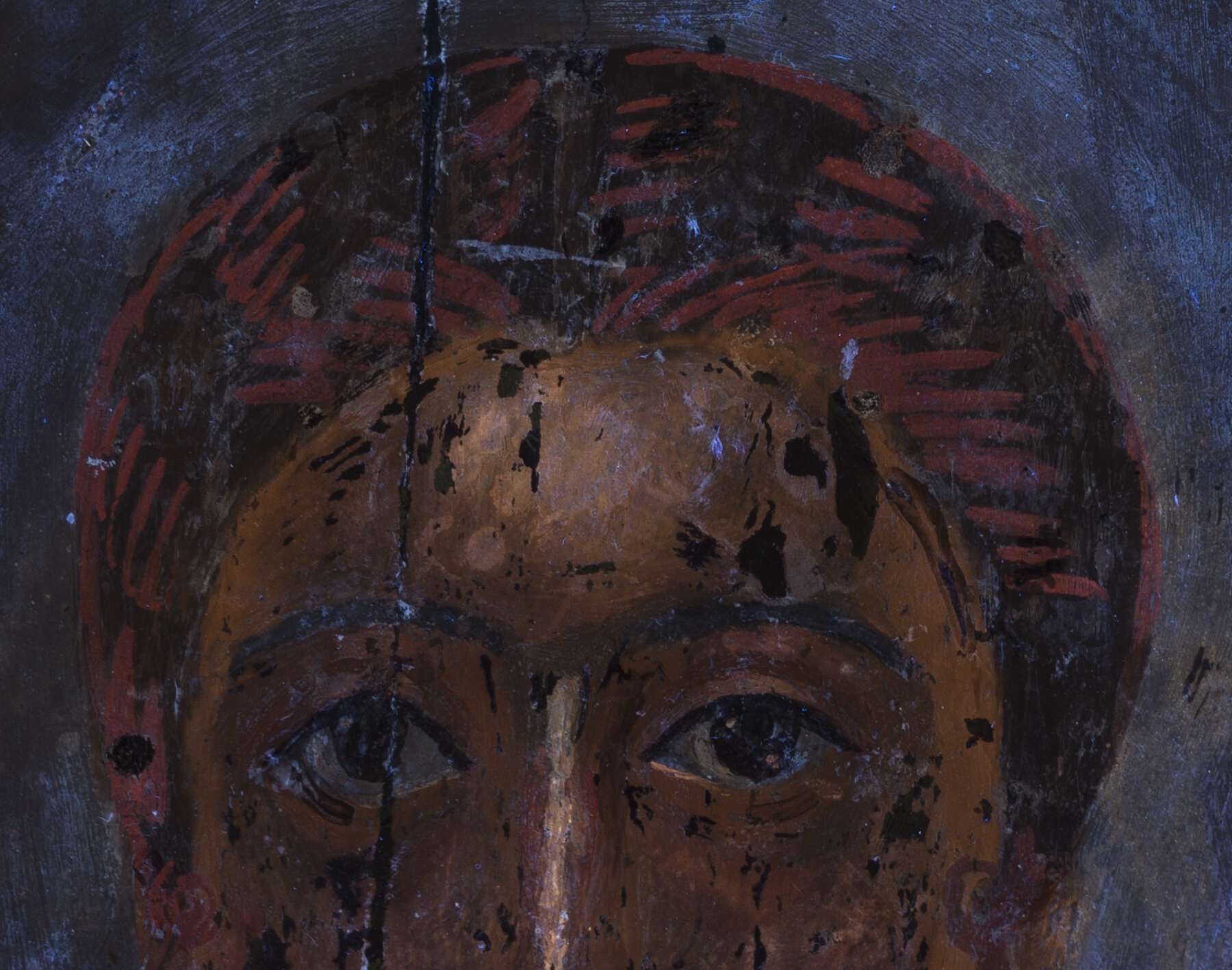 Figure 7.15
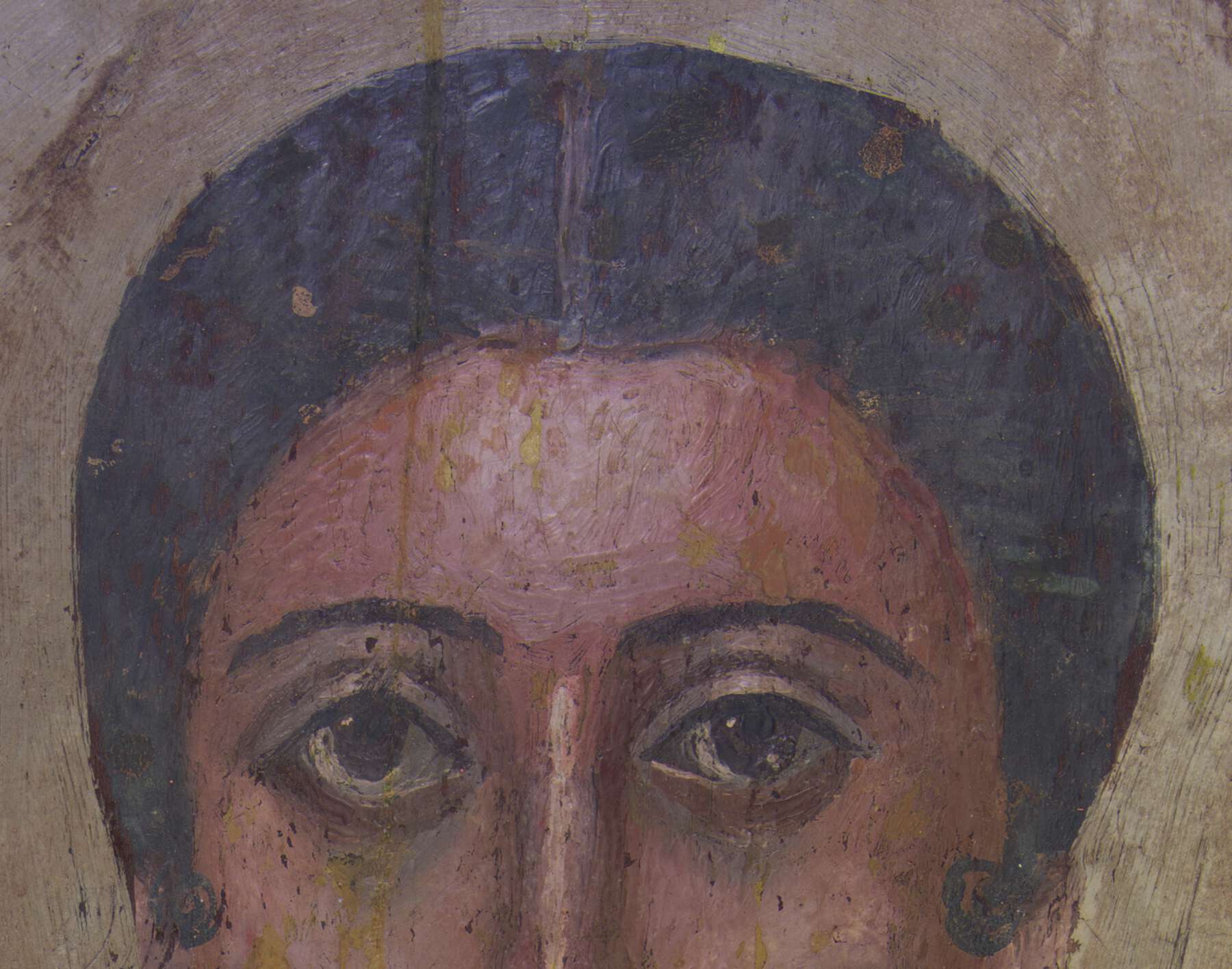 Figure 7.16
Conclusion
The setup and capture protocols developed during this investigation generate consistent MBR images using accessible tools and software. MBR is a relatively straightforward and low-tech method for characterizing and mapping materials, such as indigo, across the surface of an object, which can inform further analysis or enable extrapolation from spot assays. MBI image suites can help clarify material responses by considering behaviors across different wavelengths and bands.
Further research could advance the noninvasive characterization and mapping of materials in cultural heritage objects.16 MBR performed with the filters discussed here could be used to investigate the effectiveness of imaging indigo-containing paint that has been covered as a result of conservation treatment. Additional imaging and analysis of Romano-Egyptian portraits and reference paint-outs could address questions about how aging impacts indigo’s MBR response. Future MBR studies could investigate new combinations of filters to target other materials with pronounced changes in reflectance within the ultraviolet, visible, and infrared spectral regions.
Acknowledgments
The authors would like to thank Anna Serotta for advising and helping to carry out the imaging; Sylvana Barrett and Marie Svoboda for sharing their expertise with historical painting practices; Caroline Cartwright for generously performing wood identification on the BKM portraits; BKM curators Edward Bleiberg and Yekaterina Barbash and curatorial assistant Kathy Zurek Doule for enabling this research; BKM interns Colleen Watkins, Natasha Kung, Meredith Menache, Josephine Ren, Ariana Smith, Anneliese Holmes, and Heather Hodge for their assistance in making the paint-out boards; Nathan Griffith for sharing his understanding of image processing softwares and functions; and the BKM conservation department along with Anne Pasternak, Shelby White and Leon Levy Director of the Brooklyn Museum, for supporting this project. Raman analysis was carried out at the Department of Scientific Research of the Metropolitan Museum of Art, as part of the Network Initiative for Conservation Science (NICS). Support for NICS was provided by a grant from the Andrew W. Mellon Foundation.
Notes
- . ↩
- . ↩
- . ↩
- . ↩
- , sections 6.4.7, 6.5.8. ↩
- This Spectralon exhibits 99 percent reflectance from 400 to 1600 nanometers, decreasing to a minimum of approximately 95 percent reflectance from 250 to 400 nanometers and from 1600 to 2500 nanometers. “Spectralon® Diffuse Reflectance Standards,” Labsphere, accessed December 21, 2018, https://www.labsphere.com/site/assets/files/2628/pb-13058rev01_standards.pdf. ↩
- Indigo, Indian, powder, Indigofera tinctoria, Kremer Pigments item 36000. ↩
- Post-processing desaturation is not included in the MBR technique performed at the Smithsonian Museum Conservation Institute, which used a camera modified to capture monochrome images. The system used for this paper was based on modifications to BKM’s existing MBI kit, which does not include a monochrome camera but is more accessible. ↩
- MidOpt bandpass filter transmission curves from “BP600 Dark Red Bandpass Filter” and “BP735 Near-IR Bandpass Filter,” MidOpt, accessed January 11, 2019, http://midopt.com/filters/bp660/; and http://midopt.com/filters/bp735/. ↩
- Presentation delivered on May 16, 2019, American Institute for Conservation, 47th Annual Meeting, “Materials Characterization with Multiband Reflectance Image Subtraction at the Brooklyn Museum: A New Tool for the Multiband Imaging Kit.” ↩
- This may seem mathematically arbitrary, but it follows the optical behavior of indigo, as it absorbs at 660 nanometers and appears dark in the BP660 capture and reflects at 735 nanometers and appears light in the BP735 capture. ↩
- . ↩
Materials list:
Indigo (see note 7)
Linden wood (Tilia americana), Woodworkers Source
Cow hide glue, cubes, Kremer Pigments item 63020
Rabbit skin glue, cubes, Kremer Pigments item 63025
Rabbit skin glue, undated historical BKM lab materials
Beeswax, natural, bright yellow beads, Kremer Pigments item 62200
White beeswax, white beads, Natural Pigments
Gypsum, food-grade calcium sulfate, LD Carlson
Egyptian blue, Kremer Pigments item 10060
Red Moroccan ochre, Kremer Pigments item 116430
Orpiment, Kremer Pigments item 10700
Madder lake, made from natural roots, Kremer Pigments item 37202
Lead white, undated historical BKM lab materials
Vine black, Kremer Pigments item 47000
↩- All MBR images in this paper have been slightly enhanced for print. ↩
- The red lake pigment is most likely madder, based on the characteristic pinkish orange fluorescence observed in UVF and on the well-documented use of madder in this part of the classical world; see . ↩
- . ↩
| words